. 2018 Jul;32(7):3614-3622.
doi: 10.1096/fj.201701164R. Epub 2018 Feb 5.

A role for heat shock factor 1 in hypercapnia-induced inhibition of inflammatory cytokine expression

Affiliations

A role for heat shock factor 1 in hypercapnia-induced inhibition of inflammatory cytokine expression

Ziyan Lu et al. FASEB J. 2018 Jul.

Abstract

Hypercapnia, elevated levels of CO2 in the blood, is a known marker for poor clinical prognosis and is associated with increased mortality in patients hospitalized with both bacterial and viral pneumonias. Although studies have established a connection between elevated CO2 levels and poor pneumonia outcomes, a mechanistic basis of this association has not yet been established. We previously reported that hypercapnia inhibits expression of key NF-κB-regulated, innate immune cytokines, TNF-α, and IL-6, in LPS-stimulated macrophages in vitro and in mice during Pseudomonas pneumonia. The transcription factor heat shock factor 1 (HSF1) is important in maintaining proteostasis during stress and has been shown to negatively regulate NF-κB activity. In this study, we tested the hypothesis that HSF1 activation in response to hypercapnia results in attenuated NF-κB-regulated gene expression. We found that hypercapnia induced the protein expression and nuclear accumulation of HSF1 in primary murine alveolar macrophages and in an alveolar macrophage cell line (MH-S). In MH-S cells treated with short interfering RNA targeting Hsf1, LPS-induced IL-6 and TNF-α release were elevated during exposure to hypercapnia. Pseudomonas-infected Hsf1+/+ (wild-type) mice, maintained in a hypercapnic environment, showed lower levels of IL-6 and TNF-α in bronchoalveolar lavage fluid and IL-1β in lung tissue than did infected mice maintained in room air. In contrast, infected Hsf1+/- mice exposed to either hypercapnia or room air had similarly elevated levels of those cytokines. These results suggest that hypercapnia-mediated inhibition of NF-κB cytokine production is dependent on HSF1 expression and/or activation.-Lu, Z., Casalino-Matsuda, S. M., Nair, A., Buchbinder, A., Budinger, G. R. S., Sporn, P. H. S., Gates, K. L. A role for heat shock factor 1 in hypercapnia-induced inhibition of inflammatory cytokine expression.

Keywords: bacterial infections; lung; macrophage; rodent; stress response.


Conflict of interest statement

The authors thank Dr. Karen Ridge and Dr. Navdeep Chandel (both from Northwestern University) for advice, reagents, and technical assistance. The authors acknowledge support from the U.S. National Institutes of Health (NIH) National Heart, Lung, and Blood Institute (Grants K01 HL108860, R01 HL131745, and P01 HL071643), and NIH National Institute on Aging (Grant P01 AG049665). The authors declare no conflicts of interest.

Figures
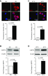
Figure 1
Figure 1
Hypercapnia and heat shock increase HSF1 protein expression and nuclear accumulation associated with increased HSP70 expression. MH-S cells were cultured in 5% CO2 [normocapnia (NC)] or 15% CO2 [hypercapnia (HC)] for 16 h. Cells were then fixed and stained for HSF1 and imaged with an IF microscope. A) HSF1 protein expression was determined via quantification of fluorescence. B) Alternatively, cells were lysed after 16 h exposure to 5 or 15% CO2 for determination of HSP70 expression in whole cell lysate. For heat shock experiments, MH-S cells were exposed to 42°C for 1 h, then allowed to recover for 1 h, or were maintained continuously at 37°C as controls. C) Cells were fixed, stained, and imaged with an IF microscope to determine HSF1 protein expression. D) HSP70 protein expression was determined via immunoblot of whole cell lysate. IF images are representative images of n ≥ 3 independent experiments. For immunoblots, bar graphs represent means ± sem, n ≥ 3 independent experiments.
Figure 2
Figure 2
Hypercapnia and heat shock exposure reduces IL-6 and TNF secretion in macrophages independent of NF-κB regulation. A) MH-S cells were cultured in 5 or 15% CO2 for 16 h, then stimulated with LPS (1 ng/ml) in normocapnia (NC) and hypercapnia (HC), respectively; after which, supernatant levels of IL-6 and TNF were determined by ELISA. B) Alternatively, cells were exposed to 42°C for 1 h, followed by recovery at 37°C for 1 h, or were maintained at 37°C as controls, then stimulated with LPS (1 ng/ml) for 6 h; after which, IL-6 and TNF release was quantified by ELISA. To determine NF-κB activation, MH-S cells exposed to 5 or 15% CO2 for 16 h, then stimulated with LPS (1 ng/ml) in NC or HC cells for 1 h. After LPS stimulation, cellular fractionation was performed to isolate cytoplasmic and nuclear compartments. C, D) IκBα (C) and RelA/p65 (D) were determined in the cytoplasmic and nuclear fractions, respectively, using immunoblot. Bar graphs represent means ± sem, n ≥ 3 independent experiments.
Figure 3
Figure 3
HSF1 is required for hypercapnia-mediated IL-6 inhibition. A) MH-S cells were treated with HSF1 siRNA or control siRNA for 48 h (at 37°, 5% CO2). Cells were then exposed to NC or HC for an additional 16 h; after which, HSF1 protein expression was determined by immunoblot; bar graphs represent means ± sem, n ≥ 3 animals. B) Alternatively, cells were treated with HSF1 or control siRNA for 72 h, exposed to normocapnia (NC) or hypercapnia (HC) for 16 h, then stimulated with LPS (1 ng/ml) for 6 h in NC or HC, respectively. IL-6 levels were determined in cellular supernatant by ELISA. Bar graphs represent means ± sem in duplicate, n ≥ 3 animals.
Figure 4
Figure 4
HSF1 and HSP70 expression is increased in hypercapnia-exposed mice. Wild-type C57BL/6 mice were exposed to normoxic hypercapnia (21% O2, 10% CO2) or air, as a control, for 3 d and then euthanized. A) HSF1 expression was determined in homogenized lung tissue by immunoblot. B) In a separate set of experiments, C57BL/6 mice were infected intratracheally with an ad-Luc1 adenovirus vector; 5 d after transfection, animals were exposed to air or normoxic hypercapnia (21% O2, 10% CO2). Mice were euthanized after exposure for 7 d to hypercapnia (or air as the control), and luciferase activity was measured in whole lung homogenates. C) C57BL/6 mice were exposed to normoxic hypercapnia (21% O2, 10% CO2), or air as control, for 3 d, Then, mice were euthanized, BAL was performed, and BAL cells were fixed, immunostained, and imaged with fluorescence microscopy to determine HSF1 and HSP70 expression. Scale bar, 10 µm. Bar graphs represent means ± sem, n ≥ 3 animals in all experiments.
Figure 5
Figure 5
HSF1 is necessary for hypercapnia-induced inhibition of IL-6 and TNF in vivo. A) HSF1 and HSP70 protein expression in lung homogenates of wild-type C57BL/6 and HSF1+/− mice was determined by immunoblot; bar graphs represent means ± sem, n ≥ 3 mice. B) Wild-type and HSF1+/− C57BL/6 mice were exposed to normoxic hypercapnia (10% CO2, 21% O2), or to air as a control, for 3 d, and were then euthanized. BAL was performed, and BAL cells fixed, immunostained, and imaged using fluorescence microscopy to quantify HSF1 and HSP70 expression. Scale bar, 10 µm. C) IL-6 and TNF protein concentration was determined in cell-free BAL fluid and IL-1β levels in lung homogenate by ELISA. Bar graphs represent means ± sem, performed in duplicates, n ≥ 3 mice.

References

    1. Mohan A., Premanand R., Reddy L. N., Rao M. H., Sharma S. K., Kamity R., Bollineni S. (2006) Clinical presentation and predictors of outcome in patients with severe acute exacerbation of chronic obstructive pulmonary disease requiring admission to intensive care unit. BMC Pulm. Med. 6, 27 - PMC - PubMed
    1. Budweiser S., Jörres R. A., Riedl T., Heinemann F., Hitzl A. P., Windisch W., Pfeifer M. (2007) Predictors of survival in COPD patients with chronic hypercapnic respiratory failure receiving noninvasive home ventilation. Chest 131, 1650–1658 - PubMed
    1. MacIntyre N., Huang Y. C. (2008) Acute exacerbations and respiratory failure in chronic obstructive pulmonary disease. Proc. Am. Thorac. Soc. 5, 530–535 - PMC - PubMed
    1. Slenter R. H., Sprooten R. T., Kotz D., Wesseling G., Wouters E. F., Rohde G. G. (2013) Predictors of 1-year mortality at hospital admission for acute exacerbations of chronic obstructive pulmonary disease. Respiration 85, 15–26 - PubMed
    1. Mancebo J. (1998) Permissive hypercapnia in ARDS. Intensive Care Med. 24, 1339–1340 - PubMed

Publication types

LinkOut - more resources