. 2018 Mar;7(3):262-270.
doi: 10.1002/sctm.17-0236. Epub 2018 Feb 5.

Digit Tip Regeneration: Merging Regeneration Biology with Regenerative Medicine

Affiliations

Digit Tip Regeneration: Merging Regeneration Biology with Regenerative Medicine

Connor P Dolan et al. Stem Cells Transl Med. 2018 Mar.

Abstract

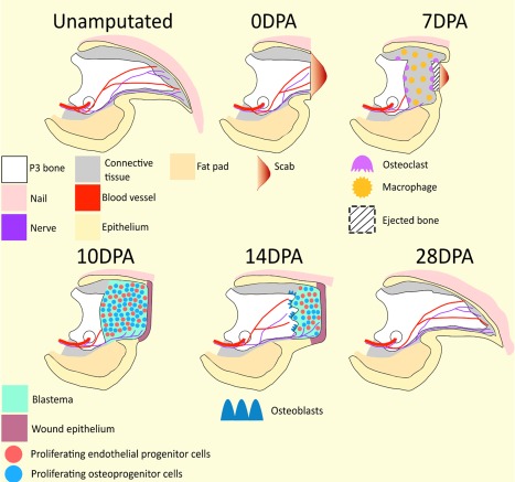
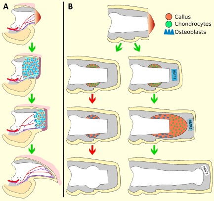
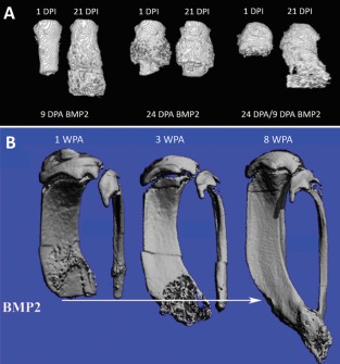
Regeneration Biology is the study of organisms with endogenous regenerative abilities, whereas Regenerative Medicine focuses on engineering solutions for human injuries that do not regenerate. While the two fields are fundamentally different in their approach, there is an obvious interface involving mammalian regeneration models. The fingertip is the only part of the human limb that is regeneration-competent and the regenerating mouse digit tip has emerged as a model to study a clinically relevant regenerative response. In this article, we discuss how studies of digit tip regeneration have identified critical components of the regenerative response, and how an understanding of endogenous regeneration can lead to expanding the regenerative capabilities of nonregenerative amputation wounds. Such studies demonstrate that regeneration-incompetent wounds can respond to treatment with individual morphogenetic agents by initiating a multi-tissue response that culminates in structural regeneration. In addition, the healing process of nonregenerative wounds are found to cycle through nonresponsive, responsive and nonresponsive phases, and we call the responsive phase the Regeneration Window. We also find the responsiveness of mature healed amputation wounds can be reactivated by reinjury, thus nonregenerated wounds retain a potential for regeneration. We propose that regeneration-incompetent injuries possess dormant regenerative potential that can be activated by targeted treatment with specific morphogenetic agents. We believe that future Regenerative Medicine-based-therapies should be designed to promote, not replace, regenerative responses. Stem Cells Translational Medicine 2018;7:262-270.


Figures

Figure 1
Figure 1
Digit tip regeneration. Unamputated: The mouse terminal phalanx (P3), is a triangular shaped cortical bone; wide at its base where it articulates with the second phalanx (P2; not pictured), gradually narrowing until it terminates as a pointed tip. Vasculature and nerves enter the P3 bone marrow via foramen referred to as os holes (shown as circles) located on either side of the ventral base of P3. The nail organ surrounds the entire digit except for the ventral surface where the ventral epidermis is an extension of the digital fat pad. Nerves and blood vessels are localized throughout the connective tissue located between the P3 bone and surrounding epidermis. 0 DPA: A scab forms in response to distal P3 amputation. Distal amputation removes 15%–20% of the P3 bone volume, but does not damage the bone marrow, fat pad, or proximal nail matrix (not shown). 7 DPA: Macrophages and other cells of the innate immune response (not shown) are scattered throughout the connective tissue and P3 bone marrow. Concurrently, large, multinucleated osteoclasts degrade the periosteal and endosteal surfaces of the P3 bone. Osteoclast activity erodes the P3 bone into two segments, thus exposing the P3 bone marrow. The remaining proximal bone stump will be reincorporated into the regenerated digit tip. 10 DPA: Epidermal migration proximal to the eroded bone functions to close the wound and eject the eroded bone. Wound closure is associated with subjacent blastema formation and the culmination of histolysis. The blastema is avascular, but is innervated. 14 DPA: Intramembranous bone redifferentiation and associated revascularization occurs proximal to distal within the wound environment. As new tissues are regenerated proximally, the distal blastema shrinks in size. 28 DPA: Digit regeneration is complete by 28 DPA, resulting in woven bone cosmetically larger than the unamputated digit. The regenerated digit tip is innervated, vascularized, and restores preamputation length. Distal is to the right. Abbreviation: DPA, days post amputation.
Figure 2
Figure 2
Schematic diagram of induced P2 regeneration. (A): The regeneration response following amputation of P3 is used as a regeneration competent model to identify factors required for blastema formation and a regeneration response. Refer to Figure 1 for full description. A number of factors, including BMP2, have been shown to be essential for the P3 regeneration response. (B): Amputation of P2 induces a dynamic wound repair response characterized by periosteal chondrogenic callus formation, followed by callus conversion to woven bone, and ultimately truncation of the bone at the original amputation plane. The wound healing response is likened to an attempt at regeneration that ultimately fails, indicated by the red arrows. BMP2 treatment to target the periosteal chondrogenic callus induces the formation of a distal chondrogenic callus that functions as a template for subsequent bone regeneration to restore the amputated skeletal length. The change in color gradient of the BMP2‐delivery‐vehicle reflects the exhaustion of BMP2. Distal is to the right.
Figure 3
Figure 3
BMP2‐induced regeneration. (A): Micro‐computed tomography (MicroCT) three‐dimensional (3D) renderings of BMP2‐treated adult mouse P2 digits. BMP2 treatment during cartilaginous peripheral callus formation, at 9 DPA, induces robust skeletal regeneration, evident by 21 DPI. BMP2‐treatment after the cartilaginous peripheral callus conversion to boney tissue at 24 DPA does not induce skeletal regeneration. Reinjury of previously healed 24 DPA P2 digits stimulates a regeneration permissive environment in which BMP2 functions to induce regeneration. (B): Sequential MicroCT 3D renderings of the BMP2‐induced hind limb regeneration response in adult mice. Amputation plane shown as arrow. Hind limbs were treated with BMP2 at 2 WPA. BMP2‐induced regeneration is evident by 3 WPA, shown as the formation of woven bone distal to the amputation plane. By 8 WPA, distal skeletal fusion is shown, indicating the regeneration response is associated with the reestablishment of skeletal patterning. (A, B) Distal is to the bottom. (A) Reprinted from Dawson et al. 11. (B) Reprinted from Yu et al. 64. Abbreviations: DPA, days post amputation; DPI, days post implantation; WPA, weeks post amputation.

References

    1. Sanchez Alvarado A. Regeneration in the metazoans: Why does it happen? Bioessays 2000;22:578–590. - PubMed
    1. Tanaka EM. The molecular and cellular choreography of appendage regeneration. Cell 2016;165:1598–1608. - PubMed
    1. Masaki H, Ide H. Regeneration potency of mouse limbs. Dev Growth Differ 2007;49:89–98. - PubMed
    1. Yu L, Han M, Yan M et al. BMP signaling induces digit regeneration in neonatal mice. Development 2010;137:551–559. - PMC - PubMed
    1. Yu L, Yan M, Simkin J et al. Angiogenesis is inhibitory for mammalian digit regeneration. Regeneration (Oxf) 2014;1:33–46. - PMC - PubMed

Publication types