. 2018 Feb 27;57(8):1306-1315.
doi: 10.1021/acs.biochem.7b01097. Epub 2018 Feb 6.

X-ray and EPR Characterization of the Auxiliary Fe-S Clusters in the Radical SAM Enzyme PqqE

Affiliations

X-ray and EPR Characterization of the Auxiliary Fe-S Clusters in the Radical SAM Enzyme PqqE

Ian Barr et al. Biochemistry. .

Abstract

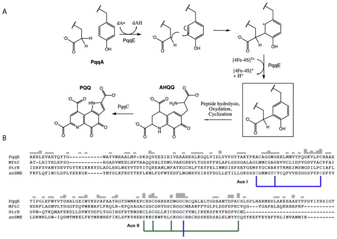
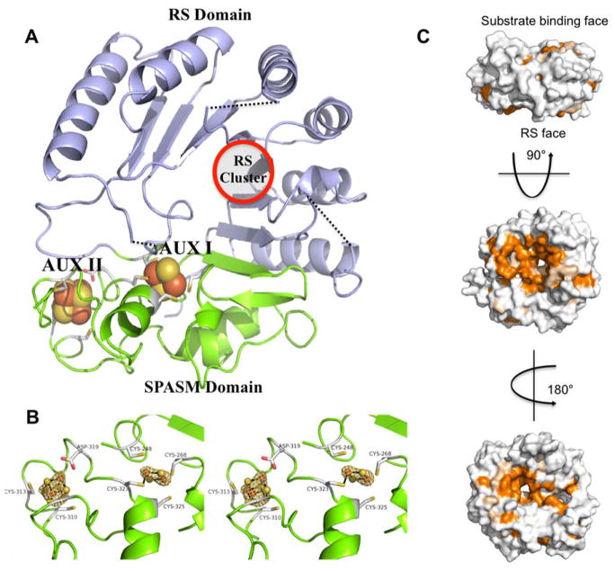
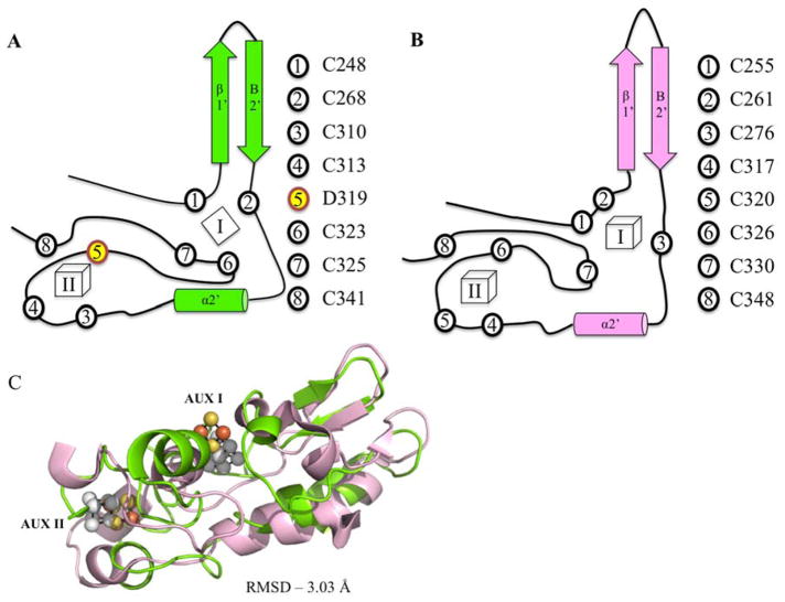
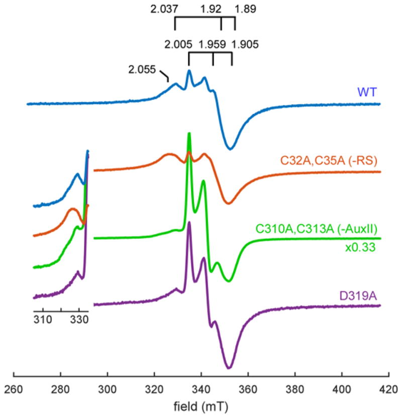
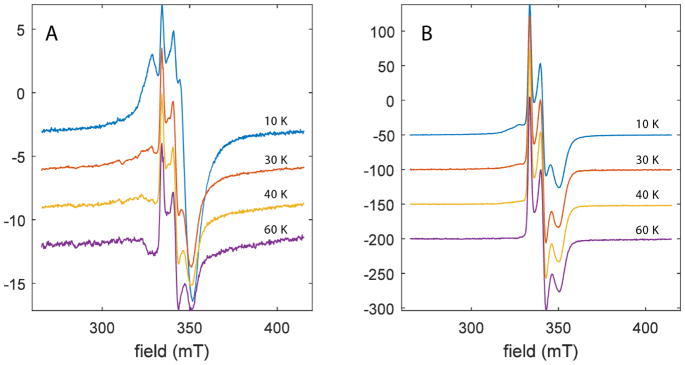
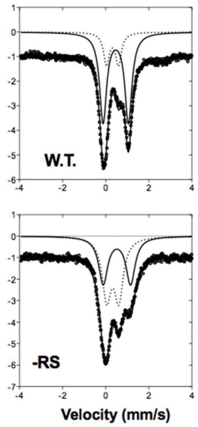
The Radical SAM (RS) enzyme PqqE catalyzes the first step in the biosynthesis of the bacterial cofactor pyrroloquinoline quinone, forming a new carbon-carbon bond between two side chains within the ribosomally synthesized peptide substrate PqqA. In addition to the active site RS 4Fe-4S cluster, PqqE is predicted to have two auxiliary Fe-S clusters, like the other members of the SPASM domain family. Here we identify these sites and examine their structure using a combination of X-ray crystallography and Mössbauer and electron paramagnetic resonance (EPR) spectroscopies. X-ray crystallography allows us to identify the ligands to each of the two auxiliary clusters at the C-terminal region of the protein. The auxiliary cluster nearest the RS site (AuxI) is in the form of a 2Fe-2S cluster ligated by four cysteines, an Fe-S center not seen previously in other SPASM domain proteins; this assignment is further supported by Mössbauer and EPR spectroscopies. The second, more remote cluster (AuxII) is a 4Fe-4S center that is ligated by three cysteine residues and one aspartate residue. In addition, we examined the roles these ligands play in catalysis by the RS and AuxII clusters using site-directed mutagenesis coupled with EPR spectroscopy. Lastly, we discuss the possible functional consequences that these unique AuxI and AuxII clusters may have in catalysis for PqqE and how these may extend to additional RS enzymes catalyzing the post-translational modification of ribosomally encoded peptides.


Conflict of interest statement

Notes

The authors declare no competing financial interest.

Figures

Figure 1
Figure 1
A, proposed mechanism for carbon-carbon bond formation in the biosynthesis of PQQ, and its relation to the rest of the biosynthetic pathway. The final product of the PqqE reaction is boxed. B, ClustalW alignment of the C-terminal region of SPASM domain family members. The cysteines ligated to the innermost auxiliary cluster (Aux I) in AnSME are identified by the blue bracket, and the outermost (AuxII) are bracketed in green. In PqqE, D319 is placed exactly where, in the other three examples, there is a conserved cysteine; the latter is seen to ligate to Aux II in the AnSME X-ray structure.
Figure 2
Figure 2
Structure of MePqqE. (A) The Radical SAM (RS) domain (lavender) contains the RS (β/α)6 partial TIM barrel. The RS cluster and canonical motif are missing from the structure, but the location of the cluster has been approximated using a superposition of the AnSMEcpe structure as a model. The SPASM domain (green) harbors two auxiliary clusters. (B) Stereo view of the MePqqE SPASM domain, which contains a [2Fe-2S] cluster ligated by four cysteines and one [4Fe-4S] cluster ligated by three cysteines and one conserved aspartate. The Fe-edge (9.686 keV) anomalous difference electron density map (gold mesh) is contoured at 4.0 RMSD. (C) Surface representation of sequence conservation as calculated by the ConSurf server [http://conseq.bioinfo.tau.ac.il/] between MePqqE and 1061 members of the PqqE family (SFLD, http://sfld.rbvi.ucsf.edu/django/) using pairwise alignment. Strictly conserved residues are shown in orange, neutral substitutions are shown in tan, and variable regions are shown in white. The view is rotated 90° about the horizontal axis (middle) and 180° about the vertical axis (bottom).
Figure 3
Figure 3
Structural similarity of MePqqE and AnSMEcpe. Secondary structure and topology of MePqqE (A) and AnSMEcpe (B) show the ligation of auxiliary clusters. AUX I and AUX II are labeled as I and II. Ligating residues are represented by circles and corresponding residue numbers are shown to the right of each topological rendering. Asp 319 is shown by number 5, yellow highlighting. Panel (C) Shows the structural alignment of the SPASM domains of MePqqE in green (Fe is orange and S is yellow) and AnSMEcpe in pink (Fe and S are grey; RMSD 3.03 Å over 78 C-alphas, sequence identity 19.23%).
Figure 4
Figure 4
Continuous-wave EPR of dithionite reduced reconstituted wild-type PqqE (100 μM), PqqE -RS (200 μM), PqqE –AuxII (200 μM), and PqqE D319A (95 μM) at 10 K. We see three major EPR signals contributing to the spectrum of reduced WT PqqE: (1) a weak signal with g1 = 2.037 which corresponds to the radical SAM cluster (see simulation in Figure S2), (2) a signal with g1 = 2.055, and (3) a signal with g1 = 2.005, g2 = 1.959, g3 = 1.905, which corresponds to the auxiliary clusters. All spectra have been scaled according to the concentration of PqqE, with the PqqE –AuxII spectrum further divided by a factor of 3 for ease of comparison. Note that disruption of the AuxII cluster induces a dramatic increase in the amount of [2Fe–2S]+ signal relative to the [4Fe–4S]+ cluster signals in most samples. Inset focuses on the g1-region for the [4Fe–4S]+ cluster signals.
Figure 5
Figure 5
Temperature dependence of EPR spectra of DTH-reduced wild-type PqqE (A) and –AuxII PqqE (B). As the temperature is elevated above 10 K, the [4Fe-4S]+ signals that are dominated by AuxII (Figure 4) completely disappear, due to fast relaxation that is a signature of [4Fe-4S]+ clusters. At 60 K, all that remains is a signal with g-values = [2.0049, 1.958, 1.906]. The average g-value = 1.9563 for the remaining signal is consistent with that measured for several all cysteinyl-coordinated, ferredoxin-type [2Fe-2S]+ clusters (Table S1).
Figure 6
Figure 6
Zero-field, 4 K Mossbauer spectra of reconstituted PqqE WT and PqqE -RS knockout show loss of 4Fe-4S clusters and presence of 2Fe-2S cluster. Data (circles) were fit to two doublets, representing [4Fe-4S] (solid line) and [2Fe-2S] (dotted line). WT spectrum from Barr et al., 2016.

Similar articles

Cited by

References

    1. Duine JA. The PQQ story. J Biosci Bioeng. 1999;88:231–236. - PubMed
    1. Barr I, Latham JA, Iavarone AT, Chantarojsiri T, Hwang JD, Klinman JP. Demonstration That the Radical S-Adenosylmethionine (SAM) Enzyme PqqE Catalyzes de Novo Carbon-Carbon Cross-linking within a Peptide Substrate PqqA in the Presence of the Peptide Chaperone PqqD. J Biol Chem. 2016;291:8877–8884. - PMC - PubMed
    1. Latham JA, Iavarone AT, Barr I, Juthani PV, Klinman JP. PqqD Is a Novel Peptide Chaperone That Forms a Ternary Complex with the Radical S-Adenosylmethionine Protein PqqE in the Pyrroloquinoline Quinone Biosynthetic Pathway. J Biol Chem. 2015;290:12908–12918. - PMC - PubMed
    1. Grell TAJ, Goldman PJ, Drennan CL. SPASM and Twitch domains in S-Adenosylmethionine (SAM) radical enzymes. J Biol Chem. 2014;290:3964–3971. - PMC - PubMed
    1. Haft DH, Basu MK. Biological systems discovery in silico: radical S-adenosylmethionine protein families and their target peptides for posttranslational modification. J Bacteriol. 2011;193:2745–2755. - PMC - PubMed

Publication types

MeSH terms

LinkOut - more resources