

Review
doi: 10.1021/acs.chemrev.7b00181.
Epub 2018 Feb 6.
Lysine Acetylation Goes Global: From Epigenetics to Metabolism and Therapeutics
Affiliations
- PMID: 29405707
- PMCID: PMC6609103
- DOI: 10.1021/acs.chemrev.7b00181
Item in Clipboard
Review
Lysine Acetylation Goes Global: From Epigenetics to Metabolism and Therapeutics
Chem Rev.
.
Abstract
Post-translational acetylation of lysine residues has emerged as a key regulatory mechanism in all eukaryotic organisms. Originally discovered in 1963 as a unique modification of histones, acetylation marks are now found on thousands of nonhistone proteins located in virtually every cellular compartment. Here we summarize key findings in the field of protein acetylation over the past 20 years with a focus on recent discoveries in nuclear, cytoplasmic, and mitochondrial compartments. Collectively, these findings have elevated protein acetylation as a major post-translational modification, underscoring its physiological relevance in gene regulation, cell signaling, metabolism, and disease.
Figures
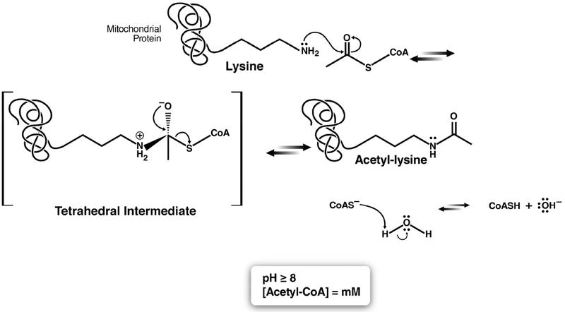
Proposed reaction mechanism of spontaneous acetylation in the mitochondria.
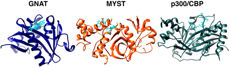
Structures of catalytic KAT domains from GNAT (human GCN5, blue, PDB: 1Z4R), MYST (human MOZ, orange, PDB: 2RC4), and KAT3A/B(CBP/p300) (human KAT3B(p300), gray, PDB: 3BIY) families. Acetyl-CoA is shown in cyan. Images rendered in Chimera (UCSF).
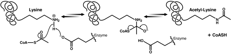
Proposed reaction mechanism for GNAT family KATs.
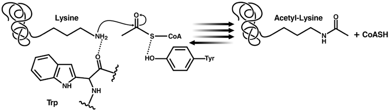
Proposed reaction mechanism for p300 family KATs.
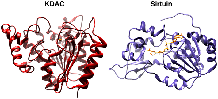
Structures of catalytic KDAC domains from KDAC (human KDAC2, red, PDB: 4LXZ) and Sirtuin (human SIRT1, purple, PDB: 4I5I families). KDAC zinc and Sirtuin NAD are shown in yellow. Images rendered in Chimera (UCSF).
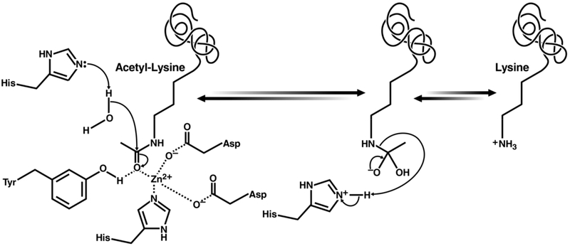
Proposed reaction mechanism for class I, II, and IV KDACs.
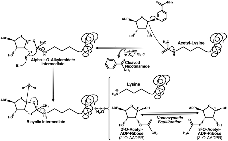
Proposed reaction mechanism for class III KDACs/sirtuins. Reprinted with permission from ref . Copyright 2010 The Royal Society of Chemistry.
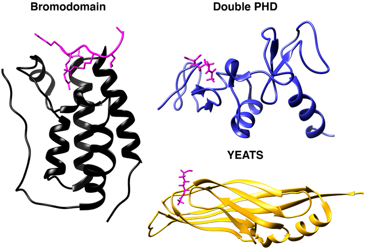
Structures of acetylation reader domains: Bromodomain (human BRD4, black, PDB: 3UVW), double PHD (human DPF3, blue, PDB: 2KWJ), and YEATS (human AF9, yellow, PDB: 4TMP). Acetyl-lysine ligands shown in pink. Images rendered in Chimera (UCSF).
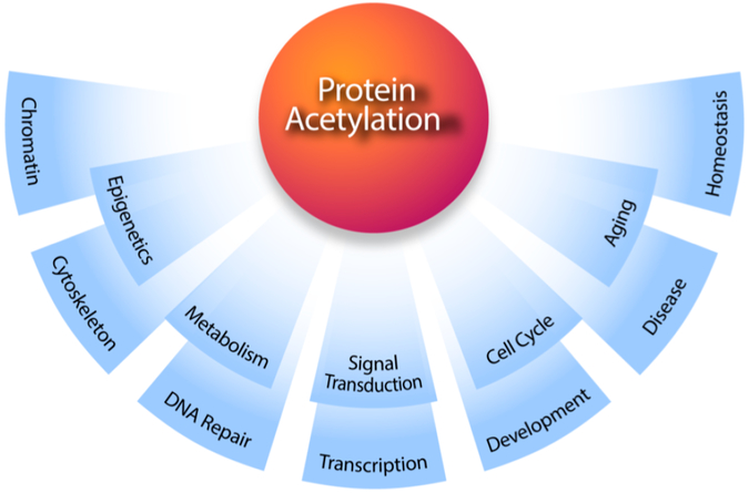
Acetylome studies reveal the scope of biological functions regulated by acetylation in mammalian cells.
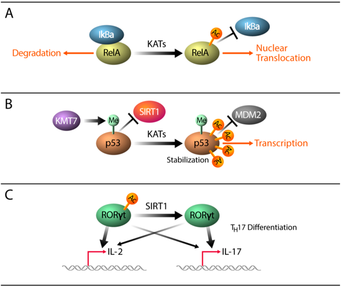
Mechanisms driving acetylation dependent regulation of transcription factors.
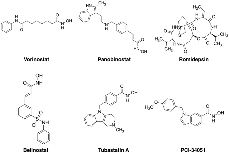
Selected chemical structures of KDAC inhibitors.
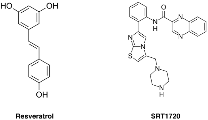
Selected chemical structures of sirtuin activators.
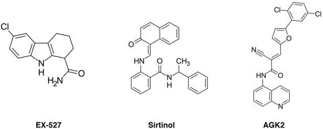
Selected chemical structures of sirtuin inhibitors.
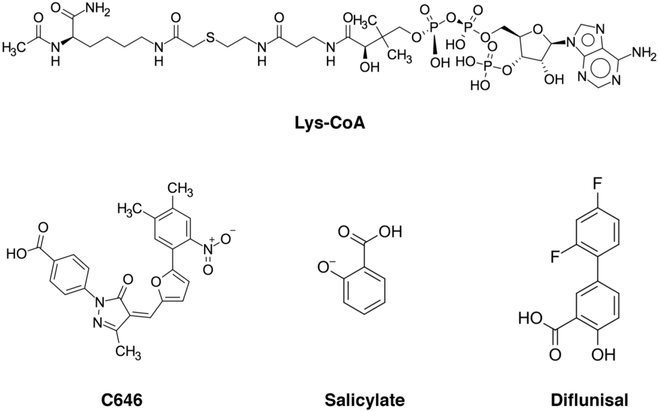
Selected chemical structures of KAT inhibitors.
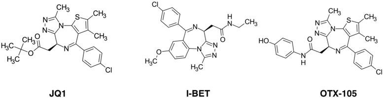
Selected chemical structures of BET inhibitors.
References
-
- Aksnes H; Drazic A; Marie M; Arnesen T First Things First: Vital Protein Marks by N-Terminal Acetyltransferases. Trends Biochem. Sci 2016, 41, 746–60. - PubMed
-
- Struhl K Histone Acetylation and Transcriptional Regulatory Mechanisms. Genes Dev. 1998, 12, 599–606. - PubMed
-
- Grunstein M Histone Acetylation in Chromatin Structure and Transcription. Nature 1997, 389, 349–52. - PubMed
-
- Strahl BD; Allis CD The Language of Covalent Histone Modifications. Nature 2000, 403, 41–5. - PubMed
-
- Turner BM Histone Acetylation and an Epigenetic Code. BioEssays 2000, 22, 836–45. - PubMed
Publication types
MeSH terms
Substances
Grants and funding
LinkOut - more resources
Full Text Sources
Other Literature Sources
Miscellaneous
