

Scorpion Venom Causes Apoptosis by Increasing Reactive Oxygen Species and Cell Cycle Arrest in MDA-MB-231 and HCT-8 Cancer Cell Lines
- PMID: 29405760
- PMCID: PMC5881405
- DOI: 10.1177/2156587217751796
Scorpion Venom Causes Apoptosis by Increasing Reactive Oxygen Species and Cell Cycle Arrest in MDA-MB-231 and HCT-8 Cancer Cell Lines
Abstract
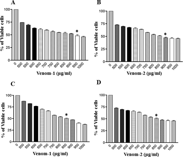
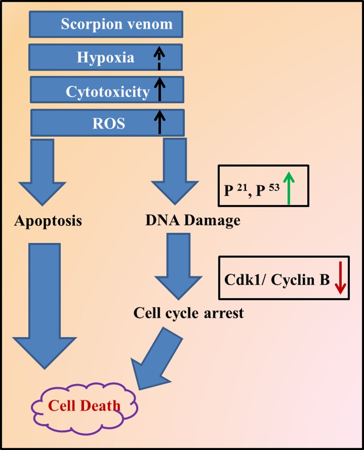
Objectives: The objective of this study was to examine the effect of scorpion venoms on cancer cell progression, apoptosis, and cell cycle arrest. Scorpion venoms are known to possess numerous bioactive compounds that act against cancer progression by inducing apoptosis. In this study, we have taken the venoms from the following 2 species of scorpion- Androctonus crassicauda and Leiurus quinquestriatus-and tested the anticancer properties of the venom against breast and colorectal cancer cell lines.
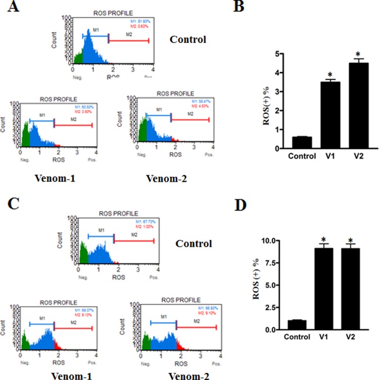
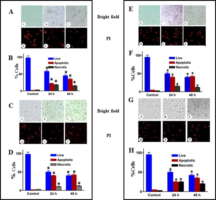
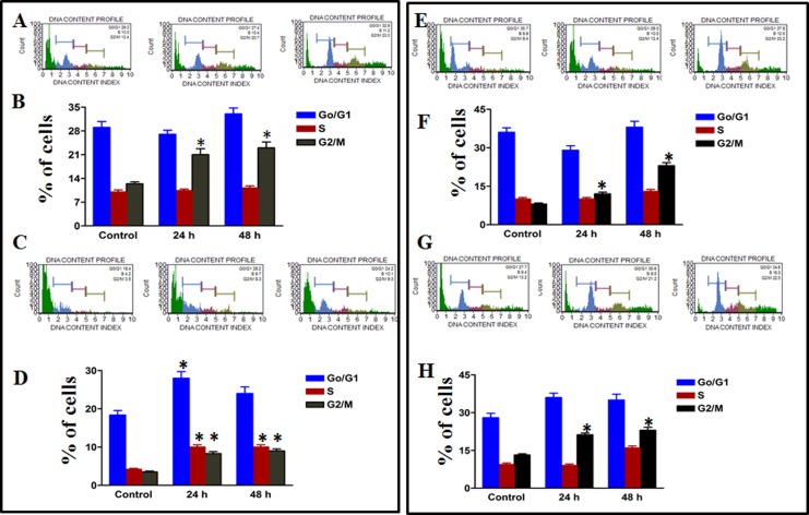
Methods: Milking of scorpion venom and culturing the breast and colorectal cancer cell lines were done according to the standard procedure. The venom cytotoxicity was assessed by MTT methods, and the cellular and nuclear changes were studied with phase contrast and propidium iodide staining, respectively. The cell cycle arrest and accumulation of reactive oxygen species were analyzed on a Muse cell analyzer.
Results: The venoms exerted cytotoxic effects on breast and colorectal cell lines in a dose- and time-dependent manner. Enhanced apoptotic cells, increase in reactive oxygen species, and cell cycle arrest were observed after challenging these cell lines with scorpion venoms.
Conclusions: Scorpion venom induces apoptosis in breast and colorectal cell lines as reflected by the changes in the cell morphology and cell cycle studies. Furthermore, a high percentage of total reactive oxygen species as well as apoptotic cells also contribute to cell death as observed after venom treatments. To the best of authors' knowledge, this is the first scientific evidence demonstrating the induction of apoptosis and cell cycle arrest by these species of scorpion venoms.
Keywords: G0/G1 phase; MTT assay; breast cancer cell lines; cell cycle assay; colorectal.
Conflict of interest statement
Figures
Similar articles
-
Scorpion (Androctonus bicolor) venom exhibits cytotoxicity and induces cell cycle arrest and apoptosis in breast and colorectal cancer cell lines.Indian J Pharmacol. 2016 Sep-Oct;48(5):537-543. doi: 10.4103/0253-7613.190742. Indian J Pharmacol. 2016. PMID: 27721540 Free PMC article.
-
In vitro determination of the efficacy of scorpion venoms as anti-cancer agents against colorectal cancer cells: a nano-liposomal delivery approach.Int J Nanomedicine. 2017 Jan 13;12:559-574. doi: 10.2147/IJN.S123514. eCollection 2017. Int J Nanomedicine. 2017. PMID: 28144138 Free PMC article.
-
Scorpion (Androctonus crassicauda) venom limits growth of transformed cells (SH-SY5Y and MCF-7) by cytotoxicity and cell cycle arrest.Exp Mol Pathol. 2011 Aug;91(1):447-54. doi: 10.1016/j.yexmp.2011.04.008. Epub 2011 Apr 22. Exp Mol Pathol. 2011. PMID: 21536027
-
Scorpion venoms as a potential source of novel cancer therapeutic compounds.Exp Biol Med (Maywood). 2014 Apr;239(4):387-93. doi: 10.1177/1535370213513991. Epub 2014 Mar 5. Exp Biol Med (Maywood). 2014. PMID: 24599885 Review.
-
Animal Venoms have Potential to Treat Cancer.Curr Top Med Chem. 2018;18(30):2555-2566. doi: 10.2174/1568026619666181221120817. Curr Top Med Chem. 2018. PMID: 30574852 Review.
Cited by
-
Superoxide Dismutase Prevents SARS-CoV-2-Induced Plasma Cell Apoptosis and Stabilizes Specific Antibody Induction.Oxid Med Cell Longev. 2022 Jan 17;2022:5397733. doi: 10.1155/2022/5397733. eCollection 2022. Oxid Med Cell Longev. 2022. PMID: 35047106 Free PMC article.
-
Monitoring the Early Antiproliferative Effect of the Analgesic-Antitumor Peptide, BmK AGAP on Breast Cancer Using Intravoxel Incoherent Motion With a Reduced Distribution of Four b-Values.Front Physiol. 2019 Jun 21;10:708. doi: 10.3389/fphys.2019.00708. eCollection 2019. Front Physiol. 2019. PMID: 31293432 Free PMC article.
-
A Novel Therapeutic Formulation for the Improved Treatment of Indian Red Scorpion (Mesobuthus tamulus) Venom-Induced Toxicity-Tested in Caenorhabditis elegans and Rodent Models.Toxins (Basel). 2023 Aug 14;15(8):504. doi: 10.3390/toxins15080504. Toxins (Basel). 2023. PMID: 37624261 Free PMC article.
-
Systematic Review of the Antitumor Activities and Mechanisms of Scorpion Venom on Human Breast Cancer Cells Lines (In Vitro Study).J Clin Med. 2025 May 4;14(9):3181. doi: 10.3390/jcm14093181. J Clin Med. 2025. PMID: 40364211 Free PMC article. Review.
-
Tityus serrulatus Scorpion Venom Induces Apoptosis in Cervical Cancer Cell Lines.Evid Based Complement Alternat Med. 2019 Jun 23;2019:5131042. doi: 10.1155/2019/5131042. eCollection 2019. Evid Based Complement Alternat Med. 2019. PMID: 31341494 Free PMC article.
References
-
- Siegel RL, Miller KD, Jemal A. Cancer statistics, 2016. CA Cancer J Clin. 2016;66:7–30. - PubMed
-
- Almutlaq BA, Almuazzi RF, Almuhayfir AA, et al. Breast Cancer in Saudi Arabia and its possible risk factors. J Cancer Policy. 2017;12:83–89.
-
- Alhazmi FG. Comparison of breast and colorectal cancer screening programs in the Netherlands and the Kingdom of Saudi Arabia. Cancer. 2016;4:157–165.
Publication types
MeSH terms
Substances
LinkOut - more resources
Full Text Sources
Other Literature Sources
Miscellaneous
