. 2018 Oct;37(10):1080-1091.
doi: 10.1177/0960327118755255. Epub 2018 Feb 6.

Diepoxybutane-induced apoptosis is mediated through the ERK1/2 pathway

Affiliations

Diepoxybutane-induced apoptosis is mediated through the ERK1/2 pathway

E Eluka-Okoludoh et al. Hum Exp Toxicol. 2018 Oct.

Abstract

Diepoxybutane (DEB) is the most potent active metabolite of butadiene, a regulated air pollutant. We previously reported the occurrence of DEB-induced, p53-dependent, mitochondrial-mediated apoptosis in human lymphoblasts. The present study investigated the role of the extracellular signal-regulated protein kinases 1 and 2 (ERK1/2) pathway in DEB-induced apoptotic signaling in exposed human lymphoblasts. Activated ERK1/2 and mitogen-activated protein (MAP) kinase/ERK1/2 kinase (MEK) levels were significantly upregulated in DEB-exposed human lymphoblasts. The MEK inhibitor PD98059 and ERK1/2 siRNA significantly inhibited apoptosis, ERK1/2 activation, as well as p53 and phospho-p53 (serine-15) levels in human lymphoblasts undergoing DEB-induced apoptosis. Collectively, these results demonstrate that DEB induces apoptotic signaling through the MEK-ERK1/2-p53 pathway in human lymphoblasts. This is the first report implicating the activation of the ERK1/2 pathway and its subsequent role in mediating DEB-induced apoptotic signaling in human lymphoblasts. These findings contribute towards the understanding of DEB toxicity, as well as the signaling pathways mediating DEB-induced apoptosis in human lymphoblasts.

Keywords: Diepoxybutane; ERK1/2; apoptosis; butadiene; extracellular signal–regulated protein kinase; p53.


Conflict of interest statement

Declaration of Conflicting Interests

The author(s) declare that there is no potential conflict of interest with respect to the research, authorship, or publication of this article.

Figures

Figure 1.
Figure 1.
DEB induces activation of the ERK pathway in exposed human lymphoblasts. TK6 lymphoblasts were exposed to 0 and 10 μM DEB for 24 and 48 h. Protein extracts (25 μg) obtained were analyzed for activation of ERK and MEK on separate blots utilizing the western blot technique. This was performed by utilizing rabbit antibodies specific for activated P-ERK (Thr202/Tyr204) or activated P-MEK (ser217/221), followed by IRDye-680 conjugated goat anti-rabbit secondary antibody. The P-ERK blots were subsequently reacted with a rabbit antibody specific for total ERK, followed by IRDye-800 conjugated goat anti-rabbit antibody. Finally, the blots were analyzed for GAPDH, a loading control; this utilized a mouse antibody and IRDye-680 conjugated goat anti-mouse secondary antibody. (a) Relative normalized P-ERK levels at each time point were obtained after quantification analysis by comparing P-ERK to total ERK ratios for each sample against the control-unexposed sample value. (b) Relative normalized P-MEK levels were obtained after quantification, followed by normalization to the corresponding GAPDH level for each sample. All quantifications were performed in triplicates, and a representative of three experiments is shown. Significance: *p < 0.05: compared to the corresponding control-unexposed cells at each time point, as well as between exposed cells at 24 h versus 48 h. DEB: diepoxybutane; GAPDH: glyceraldehyde 3-phosphate dehydrogenase; P-ERK: phospho-ERK; P-MEK: phospho-MEK.
Figure 2.
Figure 2.
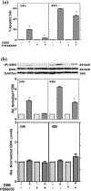
Activated ERK mediates DEB-induced apoptosis in exposed human lymphoblasts. TK6 cells were preincubated with vehicle alone or 50-μM MEK inhibitor PD98059 for 1 h. Control- and PD98059-treated cells were then exposed to 10-μM DEB for 24 and 48 h; a second set of cells was exposed to vehicle without DEB. (a) The percentage of apoptosis was determined by nuclear morphology fluorescence dye staining, as described in “Materials and methods” section. (b) Western blot analysis of activated P-ERK, total ERK (ERK), and GAPDH levels were determined using 25 μg of protein, as described in Figure 1. Relative normalized P-ERK levels for each sample were obtained after quantification analysis (in triplicates) by utilizing the ratio of P-ERK to total ERK. A representation of three experiments is shown. Significance: *p < 0.05: compared to the corresponding control-unexposed cells, as well as between DEB-exposed cells with and without the MEK inhibitor at each time point. *p < 0.05: comparison between 24 h and 48 h inhibition of apoptosis and activated ERK levels by MEK inhibitor PD98059. DEB: diepoxybutane; GAPDH: glyceraldehyde 3-phosphate dehydrogenase; P-ERK: phospho-ERK.
Figure 3.
Figure 3.
p53 is a downstream target of ERK in human lymphoblasts undergoing DEB-induced apoptosis. TK6 cells were preincubated with vehicle alone or 50-μM PD98059 for 1 h, followed by exposure to vehicle alone or 10-μM DEB for 24 or 48 h. (A) The percentage of apoptotic cells were determined by nuclear morphology fluorescence dye staining, as described in “Materials and methods” section. (b) Levels of phospho-p53 (serine-15) and GAPDH were analyzed by the western blot technique, using 25 μg of extract protein. This was accomplished by utilizing a rabbit antibody specific for phospho-p53 (serine-15) and a mouse antibody specific for GAPDH, a loading control. IRDye-680 conjugated goat anti-rabbit and IRDye-800 conjugated goat anti-mouse secondary antibodies were utilized for the detection and quantitation phospho-p53 (serine-15) and GAPDH levels, respectively. The graph shows relative normalized phospho-p53 (serine-15) levels obtained after quantification analysis (in triplicates) by normalizing each point to the corresponding GAPDH levels. A representation of three experiments is shown. Significance: *p < 0.05: compared to the corresponding control-unexposed cells, as well as between DEB-exposed cells with and without the MEK inhibitor at each time point. *p < 0.05: comparison between 24 h and 48 h inhibition of apoptosis and activated ERK levels by MEK inhibitor PD98059. DEB: diepoxybutane; GAPDH: glyceraldehyde 3-phosphate dehydrogenase.
Figure 4.
Figure 4.
ERK 1/2 siRNA knockdown of DEB-induced apoptosis in human lymphoblasts. Human TK6 lymphoblasts were transfected with 2.5-nM Silencer Select ERK siRNA or control scrambled negative control siRNA, as described in “Materials and methods” section. Control and ERK siRNA–treated cells were then exposed to control media or 10-μM DEB at 12 h post-transfection. Cells were then assayed (in triplicates) for apoptosis, P-ERK, p53, ERK, and GAPDH at 24 h post-DEB exposure. (a) The extent of apoptosis was determined using the Caspase-Glo®3/7 assay, as described in “Materials and methods” section. (b) Western blot analysis of activated P-ERK, total p53, total ERK, and GAPDH levels were determined using 15 μg of protein, as described in Figure 1. Relative normalized P-ERK levels for each sample were obtained after quantification analysis (in triplicates) by utilizing the ratio of P-ERK to total ERK. Relative normalized p53 and ERK levels were obtained after quantification analysis (in triplicates) by normalizing to GAPDH levels. A representation of three experiments is shown. Significant differences were observed between all values for DEB-exposed samples as compared to the control-unexposed samples, and between DEB-exposed cells with control siRNA versus ERK1/2 siRNA (p < 0.05). DEB: diepoxybutane; GAPDH: glyceraldehyde 3-phosphate dehydrogenase; P-ERK: phospho-ERK.

Similar articles

Cited by

References

    1. Albertini RJ, Carson ML, Kirman CR, et al. 1,3-Butadiene: II. Genotoxicity profile. Crit Rev Toxicol 2010; 40 Suppl 1: 12–73. - PubMed
    1. Albertini RJ, Carter EW, Nicklas JA, et al. 1,3-Butadiene, CML and the t(9:22) translocation: a reality check. Chem Biol Interact 2015; 241: 32–39. - PubMed
    1. Chang SC, Seneviratne UI, Wu J, et al. 1,3-Butadiene-induced adenine DNA adducts are genotoxic but only weakly mutagenic when replicated in Escherichia coli of various repair and replication backgrounds. Chem Res Toxicol 2017; 30: 1230–1239. - PMC - PubMed
    1. Elliott EG, Trinh P, Ma X, et al. Unconventional oil and gas development and risk of childhood leukemia: assessing the evidence. Sci Total Environ 2017; 576: 138–147. - PMC - PubMed
    1. National Toxicology Program. 1,3-Butadiene. National Toxicology Program, Report on Carcinogens. Research Triangle Park, NC: U.S. Department of Health and Human Services, Public Health Service. 14th ed. 016. Available at: https://ntp.niehs.nih.gov/go/roc14/.

LinkOut - more resources