. 2018 Feb:62:1-8.
doi: 10.1016/j.placenta.2017.12.002. Epub 2017 Dec 7.

Sirtuin1 is required for proper trophoblast differentiation and placental development in mice

Affiliations

Sirtuin1 is required for proper trophoblast differentiation and placental development in mice

Kanaga Arul Nambi Rajan et al. Placenta. 2018 Feb.

Abstract

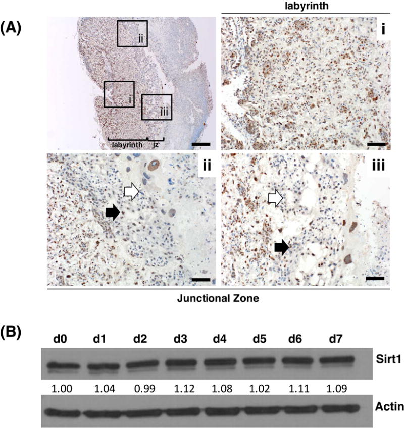
Introduction: Placental insufficiency, arising from abnormal trophoblast differentiation and function, is a major cause of fetal growth restriction. Sirtuin-1 (Sirt1) is a ubiquitously-expressed NAD-dependent protein deacetylase which plays a key role in numerous cellular processes, including cellular differentiation and metabolism. Though Sirt1 has been widely studied, its role in placentation and trophoblast differentiation is unclear.

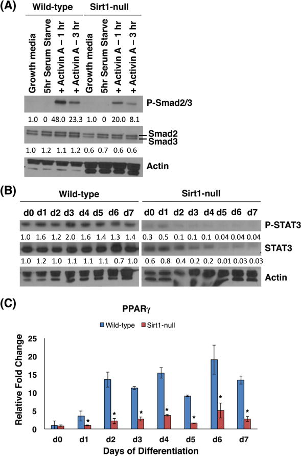
Method: Sirt1-heterozygous mice were mated and evaluated at various points during embryogenesis. In situ hybridization and immunohistochemistry were used to further characterize the placental phenotype of Sirt1-null mice. Wild-type (WT) and Sirt1-null mouse trophoblast stem cell (TSC) lines were derived from e3.5 littermate blastocysts. These cells were then evaluated at various points following differentiation. Differentiation was evaluated by expression of lineage specific markers using qPCR and flow cytometry, as well as Matrigel invasion assays. Global gene expression changes were evaluated using microarray-based RNA profiling; changes in specific pathways were validated using qPCR and western blot.

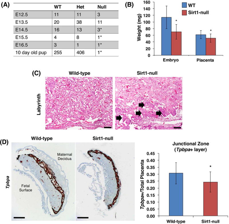
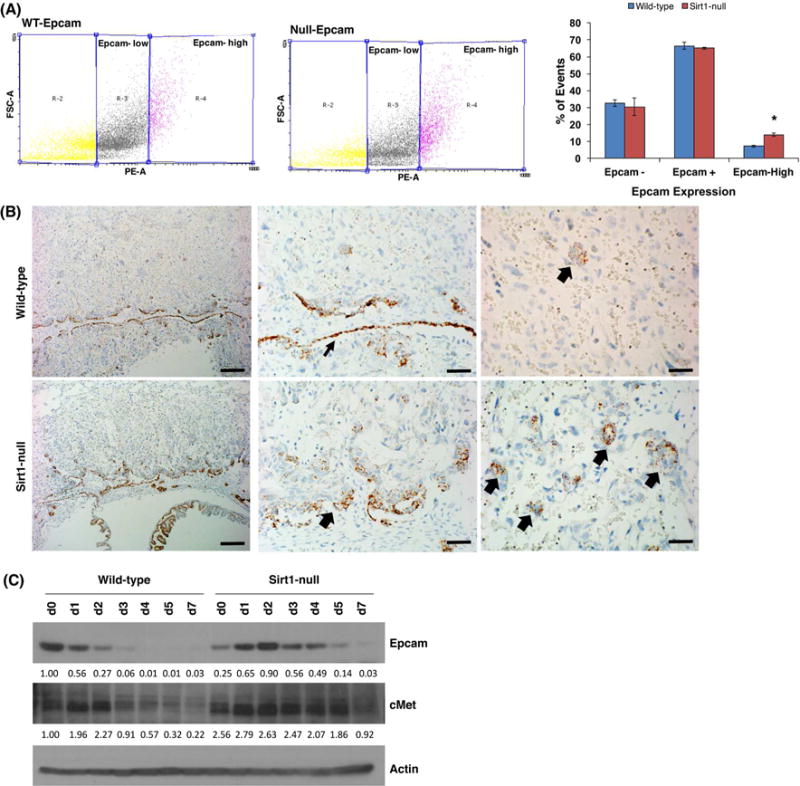
Results: In the absence of Sirt1, both embryos and placentas were small, with placentas showing abnormalities in both the labyrinthine layer and junctional zone. Sirt1-null TSCs exhibited an altered phenotype in both undifferentiated and differentiated states, phenotypes which corresponded to changes in pathways relevant to both TSC maintenance and differentiation. Specifically, Sirt1-null TSC showed blunted differentiation, and appeared to be suspended in an Epcamhigh trophoblast progenitor state.

Discussion: Our results suggest that Sirt1 is required for proper TSC differentiation and placental development.

Keywords: Differentiation; Fetal development; Fetal growth restriction; Placenta; Stem cells; Trophoblast.


Figures

Figure 1
Figure 1
Sirt1 is expressed in mouse placenta and trophoblast. (A) Sirt1 expression in E13.5 mouse placentas by immunohistochemistry, with low power image at top left, and higher power images showing (i) labyrinth, (ii) and (iii) junctional zone. Bar is 500 μm (top left) and 100 μm for (i) through (iii). Black and white block arrows mark Sirt1 positive and negative cells, respectively; jz represents the junctional zone. (B) Western blot for Sirt1 in undifferentiated (d0) and differentiated (d1 through d7) wild-type TSCs cultured in vitro. Values below the Sirt1 blot show the relative amount of the protein, normalized against actin and d0.
Figure 2
Figure 2
Absence of Sirt1 causes embryonic lethality at E13.5, with small and altered placenta. (A) Observed numbers of wild-type (WT), heterozygous (Het), and Sirt1-null (Null) embryos/pups from heterozygous matings of mice on a Sv129 background. *Indicates resorbing embryos, or pup which died soon after birth. (B) WT and null embryos and placentas were weighed after dissection at E13.5. *p=0.001 (embryo) and p = 0.0397 (placenta); n=19 WT and 10 Sirt1-null embryos. (C) H&E staining of WT and Sirt1-null E13.5 mouse placentas, showing labyrinth (Bar is 100 μm). Block arrows mark the thickened chorion in Sirt1-null labyrinth. (D) Junctional zone was evaluated by in situ hybridization using a probe against Tpbpa. Tpbpa+ area was measured and normalized to total placental area (n= 3 WT and Sirt1-null littermate pairs of embryos; p =0.0164 by paired t-test). Bar is 500 μm.
Figure 3
Figure 3
Sirt1-null mTSC lines exhibit altered proliferation and differentiation. (A) Quantification of BrdU/7AAD flow cytometry of WT and Sirt1-null TSCs in growth media. Note reduced proportion of Sirt1-null TSCs in S phase (n=3, p=0.0017). (B–D) WT and Sirt1-null TS cells differentiated in vitro over a 7-day timecourse, then subjected to qRT-PCR for lineage-specific trophoblast markers, including undifferentiated TS cells (Esrrb) (B), labyrinthine trophoblasts (Gcm1 and SynA) (C), and junctional zone (spongiotrophoblasts/Tpbpa and trophoblast giant cells/Prl3b1) (D). Values were normalized against 18S, and shown as fold change with respect to WT day 0. n=3; *p<0.01. (E) Principle Component Analysis (PCA) of microarray-based gene expression profiling data from WT and Sirt1-null TS cells at day 0, and differentiated to days 1, 5, and 7. Note separation of differentiation timepoints based on PCA1 and separation of WT and Sirt1-null cells based on PCA2. Note also the alignment of Sirt1-null day 0 TS cells with day 1-differentiated WT TS, as well as the alignment of day 7-differentiated Sirt1-null cells with day 5-differentiated WT cells. (F) Phalloidin (red) and DAPI (blue) immunofluorescent staining of differentiated (day 7) WT and Sirt1-null TS cells that have invaded Matrigel. The same number of differentiated cells (105 cells) were plated in the top chamber. Nuclei of invaded cells were manually counted, as a surrogate for cell count. n=3; *p<0.05.
Figure 4
Figure 4
Dysregulation of signaling pathways in Sirt1-null trophoblasts. (A) Western blot of phosphorylated Smad2/3, total Smad2/3 and actin in WT and Sirt1-null cells serum-starved for 5 hours and then treated with 10μg/ml Activin A. Values below the blots show the amount of protein, relative to wild-type cells in growth media only. (B) Western blot of phosphorylated Stat3, total Stat3 and Actin during WT and Sirt1-null TS cell differentiation. Values below the blots show the amount of protein, relative to wild-type cells at day 0. (C) qRT-PCR of PPARγRNA expression during WT and Sirt1-null TS cell differentiation. Data is normalized to 18S and shown with respect to WT day 0. n=3; *p<0.05.
Figure 5
Figure 5
In the absence of Sirt1, cMet-dependent Epcamhigh trophoblast progenitors are expanded. (A) Flow cytometry for Epcam detected populations of trophoblasts expressing high levels of Epcam in differentiated trophoblasts (day 5). Percentage of cells that were Epcam negative, positive, or Epcamhigh were quantified. n=3; p = 0.0003. (B) In situ hybridization for Epcam in WT and Sirt1-null cells. Note the more abundant Epcam+ cells both in the thickened chorionic plate (middle panel, block arrow; WT chorionic plate indicated by thinner block arrow) and in numerous clumps (right panel, block arrows) within the labyrinth of Sirt1-null placentas. Bar is 200 μm (left panels); 50 μm (middle panels); 35 μm (right panels). (C) Western blot of Epcam, cMet, and actin during WT and Sirt1-null TS cell differentiation. Values below the blots show the amount of protein, normalized to actin and WT d0.

Similar articles

Cited by

References

    1. Guttmacher AE, Maddox YT, Spong CY. The Human Placenta Project: placental structure, development, and function in real time. Placenta. 2014;35:303–304. - PMC - PubMed
    1. Burton GJ, Jauniaux E. What is the placenta? Am J Obstet Gynecol. 2015;213:S6–8. - PubMed
    1. Fisher SJ. Why is placentation abnormal in preeclampsia? Am J Obstet Gynecol. 2015;213:S115–S122. - PMC - PubMed
    1. Leibiger IB, Berggren PO. Sirt1: a metabolic master switch that modulates lifespan. Nat Med. 2006;12:34–36. - PubMed
    1. Michan S, Sinclair D. Sirtuins in mammals: insights into their biological function. Biochem J. 2007;404:1–13. - PMC - PubMed

Publication types