. 2018 Mar 15:386:4-11.
doi: 10.1016/j.jns.2017.12.038. Epub 2017 Dec 29.

Experimental sheep BSE prions generate the vCJD phenotype when serially passaged in transgenic mice expressing human prion protein

Affiliations

Experimental sheep BSE prions generate the vCJD phenotype when serially passaged in transgenic mice expressing human prion protein

Susan Joiner et al. J Neurol Sci. .

Abstract

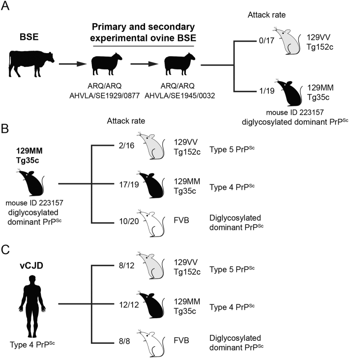
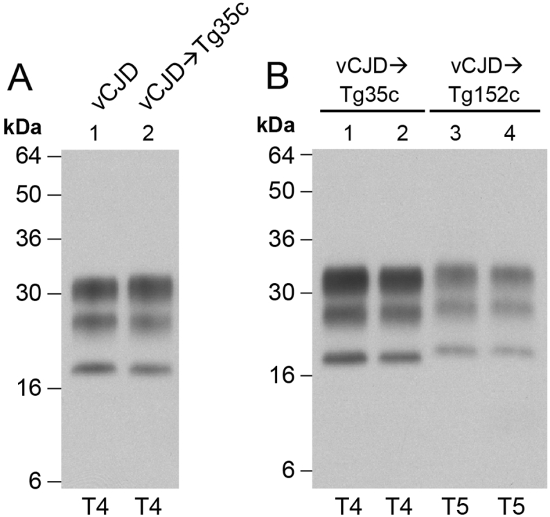
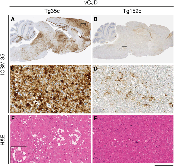
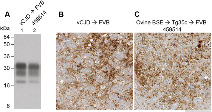
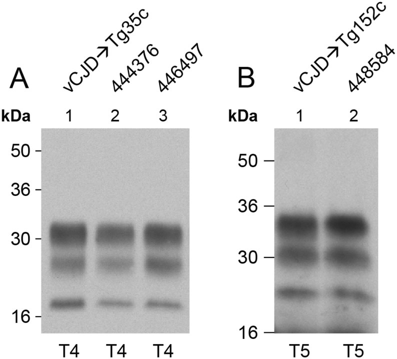
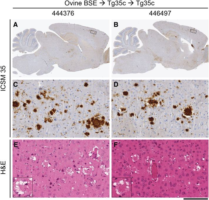
The epizootic prion disease of cattle, bovine spongiform encephalopathy (BSE), causes variant Creutzfeldt-Jakob disease (vCJD) in humans following dietary exposure. While it is assumed that all cases of vCJD attributed to a dietary aetiology are related to cattle BSE, sheep and goats are susceptible to experimental oral challenge with cattle BSE prions and farmed animals in the UK were undoubtedly exposed to BSE-contaminated meat and bone meal during the late 1980s and early 1990s. Although no natural field cases of sheep BSE have been identified, it cannot be excluded that some BSE-infected sheep might have entered the European human food chain. Evaluation of the zoonotic potential of sheep BSE prions has been addressed by examining the transmission properties of experimental brain isolates in transgenic mice that express human prion protein, however to-date there have been relatively few studies. Here we report that serial passage of experimental sheep BSE prions in transgenic mice expressing human prion protein with methionine at residue 129 produces the vCJD phenotype that mirrors that seen when the same mice are challenged with vCJD prions from patient brain. These findings are congruent with those reported previously by another laboratory, and thereby strongly reinforce the view that sheep BSE prions could have acted as a causal agent of vCJD within Europe.

Keywords: Bovine spongiform encephalopathy (BSE); Prion disease; Prions; Sheep-BSE; Transmissible spongiform encephalopathy (TSE); Variant Creutzfeldt-Jakob disease (vCJD).


Figures

Fig. 1
Fig. 1
Summary of prion transmission rates to transgenic or wild-type mice. Mice were intracerebrally inoculated with 1% (w/v) brain homogenate. Attack rates report the total of clinically affected and subclinically infected mice as a proportion of the number of inoculated mice after prolonged post-inoculation periods. The type of PrPSc observed in the brain of affected mice is also reported. (A) Serial passage of cattle BSE prions in sheep (homozygous for ovine PrP with an ARQ genotype at codons 136, 154, 171) followed by transmission of sheep BSE prions to transgenic mice homozygous for human PrP with either methionine (M) or valine (V) at residue 129. These transmissions identified a single subclinically infected 129MM Tg35c mouse (ID 223157) that propagated a diglycosylated dominant PrPSc type in brain . (B) Transmission of prions from mouse ID 223157 to further transgenic mice and to wild-type FVB mice. (C) Primary transmission of vCJD prions from patient brain (I336) to transgenic mice and to wild-type FVB mice. Full details of the transmissions shown in panels B and C are provided in the Tables.
Fig. 2
Fig. 2
Molecular strain typing of vCJD prion transmissions from patient brain to transgenic mice. (A, B) Immunoblots of proteinase-K digested 10% (w/v) brain homogenates from a vCJD patient brain or vCJD prion-inoculated transgenic mice analysed by enhanced chemiluminescence with anti-PrP monoclonal antibody 3F4. The volumes of samples loaded were varied to give comparable levels of total PrP signal intensity in each lane. (A) vCJD patient brain and a clinically affected vCJD prion-inoculated 129MM Tg35c mouse that was culled 664 days post-inoculation demonstrating faithful propagation of type 4 PrPSc (T4). (B) Lanes 1 and 2, vCJD prion-inoculated 129MM Tg35c mouse brain from two clinically affected mice (culled 688 and 664 days post inoculation) showing propagation of type 4 PrPSc (T4) compared to brain from two subclinically infected vCJD prion-inoculated 129VV Tg152c mice (culled 860 and 716 days post-inoculation) propagating type 5 PrPSc (T5). Immunohistochemical analyses of brain from the same mice shown in panel B lanes 2 and 4 are presented in Fig. 3.
Fig. 3
Fig. 3
Neuropathological analyses of vCJD prion transmissions from patient brain to transgenic mice. Panels A, C, and E, a clinically affected vCJD prion-inoculated 129MM Tg35c mouse propagating type 4 PrPSc (see Fig. 2B, lane 2) culled 664 days post-inoculation. Panels B, D, and F, a subclinically infected vCJD prion-inoculated 129VV Tg152c mouse propagating type 5 PrPSc (see Fig. 2B, lane 4) culled 716 days post-inoculation. (A, B) Sagittal sections of whole brain. (C–F) Higher power magnification of the boxed regions shown in panels A and B; (C) cortex, (D) midbrain. (A–D) Abnormal PrP immunoreactivity stained with anti-PrP monoclonal antibody ICSM 35. (E, F) Haematoxylin- and eosin-stained sections (H&E) showing spongiform neurodegeneration including florid plaques in vCJD prion-inoculated 129MM Tg35c mouse brain (inset). Scale bars: A and B, 2 mm, C–F main panels 100 μm, inset panel E, 50 μm.
Fig. 4
Fig. 4
Immunoblot and immunohistochemical analyses of prion transmissions to wild-type FVB/N mice. (A) Immunoblot of proteinase-K digested 10% (w/v) brain homogenates analysed by enhanced chemiluminescence with anti-PrP monoclonal antibody ICSM 35. Lane 1, clinically affected FVB/N mouse inoculated with vCJD prions from patient brain culled 253 days post-inoculation. Lane 2, brain from a clinically affected FVB/N mouse (ID 459514) inoculated with 129MM Tg35c-passaged ovine BSE prions culled 603 days post-inoculation. (B, C) Deposition of abnormal PrP in the midbrain of clinically affected FVB/N mice inoculated with either vCJD prions from patient brain (B) or 129MM Tg35c-passaged ovine BSE prions (C) stained with anti-PrP monoclonal antibody ICSM 35. Scale bar; 100 μm.
Fig. 5
Fig. 5
Molecular strain typing of 129MM Tg35c-passaged ovine BSE prion transmissions to transgenic mice. (A, B) Immunoblots of proteinase-K digested 10% (w/v) brain homogenates analysed by enhanced chemiluminescence with anti-PrP monoclonal antibody 3F4. The volumes of samples loaded were varied to give roughly equivalent levels of total PrP signal intensity in each lane. (A) Brain from a clinically affected 129MM Tg35c mouse inoculated with vCJD prions from patient brain (culled 664 days post-inoculation) is compared with brain from two subclinically affected 129MM Tg35c mice inoculated with 129MM Tg35c-passaged ovine BSE prions (ID numbers 444376 and 446497, both culled 701 days post-inoculation). All three brain samples show the propagation of type 4 PrPSc (T4). Immunohistochemical analyses of brain from the same mice shown in lanes 2 and 3 are presented in Fig. 6. (B) Brain from a subclinically infected 129VV Tg152c mouse inoculated with vCJD prions from patient brain (culled 687 days post-inoculation) is compared to brain from a clinically affected 129VV Tg152c mouse (ID number 448584) inoculated with 129MM Tg35c-passaged ovine BSE prions (culled 590 days post-inoculation). Both brain samples show propagation of type 5 PrPSc (T5).
Fig. 6
Fig. 6
Neuropathological analysis of 129MM Tg35c-passaged ovine BSE prion transmissions to further 129MM Tg35c mice. Images show brain from two subclinically infected 129MM Tg35c mice inoculated with 129MM Tg35c-passaged ovine BSE prions; mouse ID numbers 444376 (panels A, C and E) and 446497 (panels B, D and F). Both mice propagated type 4 PrPSc (see Fig. 5A lanes 2 and 3) and were culled 701 days post-inoculation. (A, B) Sagittal sections of whole brain. (C–F) Higher power magnification of cortex from the boxed regions shown in panels A and B. (A–D) Abnormal PrP immunoreactivity stained with anti-PrP monoclonal antibody ICSM 35. (E, F) Haematoxylin- and eosin-stained sections (H&E) showing spongiform neurodegeneration including florid plaques (insets). Scale bars: A and B, 2 mm, C-F main panels 100 μm, inset in panels E and F, 50 μm.

Similar articles

Cited by

References

    1. Collinge J., Sidle K.C., Meads J., Ironside J., Hill A.F. Molecular analysis of prion strain variation and the aetiology of 'new variant' CJD. Nature. 1996;383:685–690. - PubMed
    1. Hill A.F., Desbruslais M., Joiner S., Sidle K.C.L., Gowland I., Collinge J. The same prion strain causes vCJD and BSE. Nature. 1997;389:448–450. - PubMed
    1. Bruce M.E. Transmissions to mice indicate that 'new variant' CJD is caused by the BSE agent. Nature. 1997;389:498–501. - PubMed
    1. Asante E.A. BSE prions propagate as either variant CJD-like or sporadic CJD-like prion strains in transgenic mice expressing human prion protein. EMBO J. 2002;21:6358–6366. - PMC - PubMed
    1. Collinge J. Variant Creutzfeldt-Jakob disease. Lancet. 1999;354:317–323. - PubMed

Publication types