

doi: 10.1016/j.nlm.2018.02.001.
Epub 2018 Feb 3.
Paradoxical accentuation of motivation following accumbens-pallidum disconnection
Affiliations
- PMID: 29408054
- PMCID: PMC5864546
- DOI: 10.1016/j.nlm.2018.02.001
Item in Clipboard
Paradoxical accentuation of motivation following accumbens-pallidum disconnection
Neurobiol Learn Mem.
2018 Mar.
Abstract
The nucleus accumbens (NAc) and ventral pallidum (VP) are reciprocally connected, and activity within this circuit is thought to promote reward learning. Inconsistent with this notion, we find that disconnecting NAc medial shell and VP greatly enhances the attribution of value to a cue that is paired with reward. This result suggests that medial NAc shell and VP are both needed for attributing value to cues yet can also oppose one-another's functional contribution.
Keywords: Nucleus accumbens; Sign-tracking; Ventral pallidum.
Copyright © 2018 Elsevier Inc. All rights reserved.
Conflict of interest statement
Figures
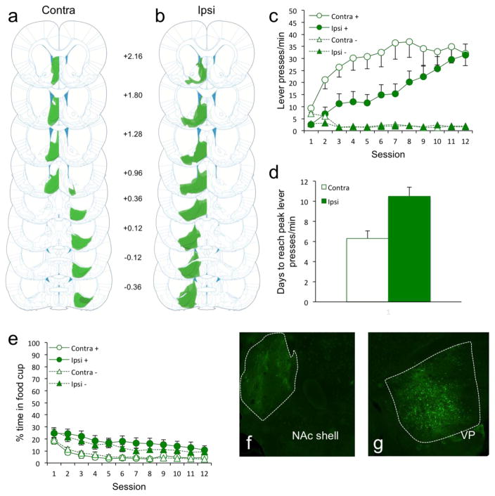
Figure 1(a–b) AAV8-hSyn-hM4Di-mCitrine cell expression in all rats of Group Contra (a; n = 14) and Group Ipsi (b; n =12). Darker areas indicate common areas of overlap in terms of expression between rats. (c) Disconnection of NAc shell and VP dramatically enhanced the acquisition of sign-tracking. (d) Group Contra rats reached peak levels of sign-tracking 40% (4 days) faster compared to Group Ipsi rats. (e) Group Contra rats spent less time in the food cup overall compared to Group Ipsi rats. (f–g) Representative brain slice showing the area of AAV8-hSyn-hM4Di-mCitrine cell expression in the NAc shell (f) and VP (g).
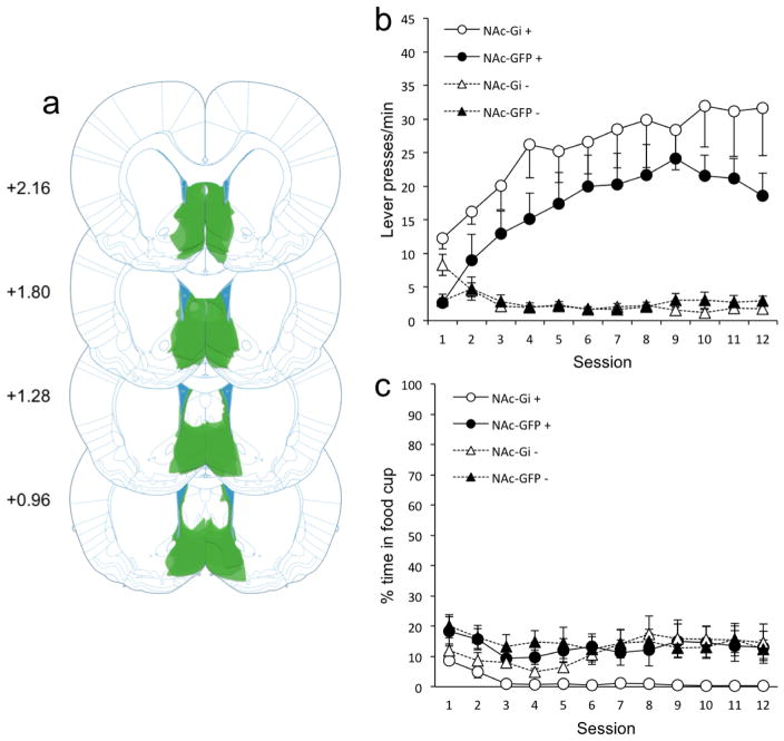
Figure 2(a) AAV8-hSyn-hM4Di-mCitrine cell expression in all Group NAc-Gi rats (n = 7). Darker areas indicate common areas of overlap in terms of expression between rats. (b) Bilateral NAc shell inhibition had no effect on sign-tracking. (c) Bilateral NAc shell inhibition lowered food cup behavior non-discriminately.
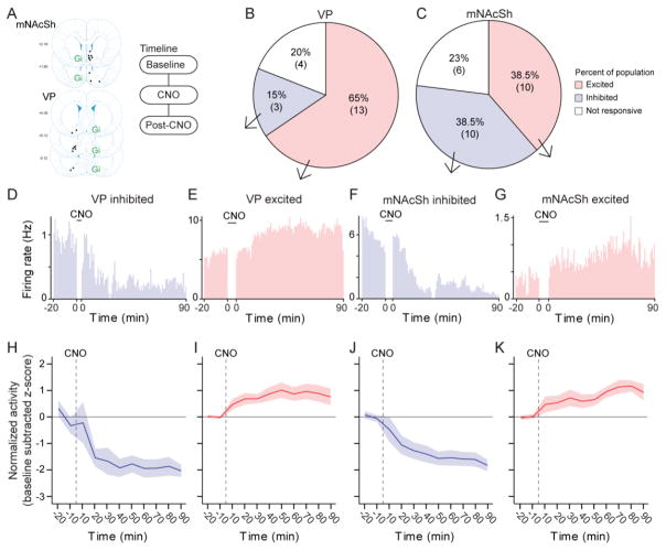
Figure 3(a) Map of tetrode placements contralateral to hM4D(Gi) infusions, and recording timeline. (b–c) Populations of units that were excited, inhibited, or unaffected by CNO from recordings in the VP (t6=2.5, p = 0.047 population size comparison) and NAc (t6=0, p = 1). Number of units is shown below percentages (n = 3 rats; n = 26 NAc units, n = 20 VP units). (d–g) Real-time histograms (1-min bins; time zero = CNO) showing single units that were excited or inhibited in the VP and NAc. (h–k) Normalized firing rates (baseline-subtracted z-score) of pooled units with inhibition or excitation in NAc and VP.
Similar articles
-
Ventral pallidum encodes relative reward value earlier and more robustly than nucleus accumbens.Nat Commun. 2018 Oct 19;9(1):4350. doi: 10.1038/s41467-018-06849-z. Nat Commun. 2018. PMID: 30341305 Free PMC article.
-
Circuit directionality for motivation: Lateral accumbens-pallidum, but not pallidum-accumbens, connections regulate motivational attraction to reward cues.Neurobiol Learn Mem. 2019 Jul;162:23-35. doi: 10.1016/j.nlm.2019.05.001. Epub 2019 May 13. Neurobiol Learn Mem. 2019. PMID: 31096040 Free PMC article.
-
Ventral Pallidum Neurons Encode Incentive Value and Promote Cue-Elicited Instrumental Actions.Neuron. 2016 Jun 15;90(6):1165-1173. doi: 10.1016/j.neuron.2016.04.037. Epub 2016 May 26. Neuron. 2016. PMID: 27238868 Free PMC article.
-
The ventral pallidum: Subregion-specific functional anatomy and roles in motivated behaviors.Prog Neurobiol. 2015 Jul;130:29-70. doi: 10.1016/j.pneurobio.2015.03.005. Epub 2015 Apr 6. Prog Neurobiol. 2015. PMID: 25857550 Free PMC article. Review.
-
Ventral pallidal regulation of motivated behaviors and reinforcement.Front Neural Circuits. 2023 Feb 2;17:1086053. doi: 10.3389/fncir.2023.1086053. eCollection 2023. Front Neural Circuits. 2023. PMID: 36817646 Free PMC article. Review.
Cited by
-
Ventral pallidum encodes relative reward value earlier and more robustly than nucleus accumbens.Nat Commun. 2018 Oct 19;9(1):4350. doi: 10.1038/s41467-018-06849-z. Nat Commun. 2018. PMID: 30341305 Free PMC article.
-
'Liking' and 'wanting' in eating and food reward: Brain mechanisms and clinical implications.Physiol Behav. 2020 Dec 1;227:113152. doi: 10.1016/j.physbeh.2020.113152. Epub 2020 Aug 23. Physiol Behav. 2020. PMID: 32846152 Free PMC article. Review.
-
Sign-tracking behavior is sensitive to outcome devaluation in a devaluation context-dependent manner: implications for analyzing habitual behavior.Learn Mem. 2020 Mar 16;27(4):136-149. doi: 10.1101/lm.051144.119. Print 2020 Apr. Learn Mem. 2020. PMID: 32179656 Free PMC article.
-
Distinct role of nucleus accumbens D2-MSN projections to ventral pallidum in different phases of motivated behavior.Cell Rep. 2022 Feb 15;38(7):110380. doi: 10.1016/j.celrep.2022.110380. Cell Rep. 2022. PMID: 35172164 Free PMC article.
-
Circuit directionality for motivation: Lateral accumbens-pallidum, but not pallidum-accumbens, connections regulate motivational attraction to reward cues.Neurobiol Learn Mem. 2019 Jul;162:23-35. doi: 10.1016/j.nlm.2019.05.001. Epub 2019 May 13. Neurobiol Learn Mem. 2019. PMID: 31096040 Free PMC article.
References
-
- Berridge KC. Motivation concepts in behavioral neuroscience. Physiology & Behavior. 2004;81:179–209. - PubMed
Publication types
MeSH terms
Grants and funding
LinkOut - more resources
Full Text Sources
Other Literature Sources
