Review
. 2018 Feb 5;44(3):284-296.
doi: 10.1016/j.devcel.2018.01.009. Epub 2018 Feb 5.

Cell Extrusion: A Stress-Responsive Force for Good or Evil in Epithelial Homeostasis

Affiliations
Review

Cell Extrusion: A Stress-Responsive Force for Good or Evil in Epithelial Homeostasis

Shizue Ohsawa et al. Dev Cell. .

Erratum in

Abstract

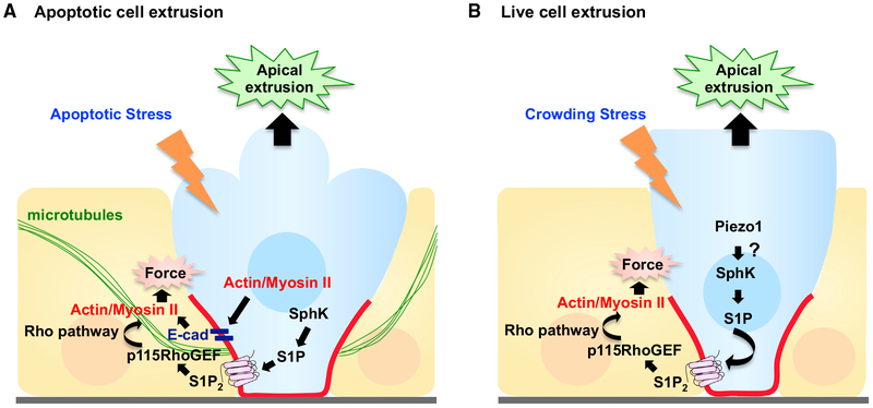
Epithelial tissues robustly respond to internal and external stressors via dynamic cellular rearrangements. Cell extrusion acts as a key regulator of epithelial homeostasis by removing apoptotic cells, orchestrating morphogenesis, and mediating competitive cellular battles during tumorigenesis. Here, we delineate the diverse functions of cell extrusion during development and disease. We emphasize the expanding role for apoptotic cell extrusion in exerting morphogenetic forces, as well as the strong intersection of cell extrusion with cell competition, a homeostatic mechanism that eliminates aberrant or unfit cells. While cell competition and extrusion can exert potent, tumor-suppressive effects, dysregulation of either critical homeostatic program can fuel cancer progression.

Keywords: apoptosis; cancer; cell competition; cell extrusion; development; epithelia; epithelial-mesenchymal transition; morphogenesis; tissue homeostasis.


Figures

Figure 1.
Figure 1.. Model for Apoptotic and Live Cell Extrusion
(A) In response to apoptotic stress, cells undergoing apoptosis produce sphingosine-1-phosphate (S1P) via sphingosine kinase (SphK), which binds to the S1P receptor (S1P2) in neighboring cells. S1P2 activates Rho signaling through p115 RhoGEF recruited basally by microtubules, triggering basal actomyosin contraction and subsequent apical extrusion of the dying cell. For simplicity, the autonomous actomyosin force also required for apoptotic cell extrusion has been omitted (see text). (B) In response to crowding stress, Piezo1 is activated, which triggers live cell extrusion. S1P-Rho signaling is again required for extrusion, but how and if Piezo1 cooperates with S1P-Rho signaling remains unclear.
Figure 2.
Figure 2.. Cell Extrusion Coupled to Drosophila Neuroblast Division and Differentiation
Proper extrusion of Drosophila neuroblasts (NBs) enables asymmetric NB divisions, daughter cell fate decision, and correct neurogenesis. Bazooka (PAR3, Baz) localizes apically in the mother epithelium, a polarity that critically is inherited by extruding NBs. During NB delamination, Inscuteable (Insc) is expressed, localizes apically, and recruits Pins. During mitotic division, several proteins (including Numb) localize to the basal cortex. This correct, asymmetric segregation of key proteins governs daughter cell fates and generates a new neuroblast and a ganglion mother cell (GMC), which produces neurons and glia.
Figure 3.
Figure 3.. Leg Folding by Extruding, Apoptotic Cell Forces
During Drosophila leg-joint development, extruding apoptotic cells (blue) pull their neighbors toward the basal side at the presumptive joint area by accumulating myosin II along their apical-basal axis. Neighboring cells accumulate myosin II at their apical surface, which triggers apical constriction and subsequent fold formation.
Figure 4.
Figure 4.. Tumor Cell Extrusion through Slit-Robo2-Ena Signaling in Cell Competition
(A) Polarity-deficient loser scrib cells (blue loser cells) are predominantly extruded basally from Drosophila epithelial tissue when confronted with wild-type, winner cells (orange winner cells). In polarity-deficient cells, JNK signaling activates Slit-Robo2-Ena signaling, which dysregulates E-cadherin to drive basal extrusion. (B) In the absence of Slit-Robo2-Ena signaling, polarity-deficient loser cells (blue) escape from extrusion and overgrow in the epithelial tissue. (C) Hyperactivation of Slit-Robo2-Ena signaling results in hyperextrusion and luminal tumors.
Figure 5.
Figure 5.. Extrusion Direction Impacts Cancer Cell Outcome
Epithelial tissues (orange) remove either live or dying cells (blue) in response to crowding or apoptotic stimuli, respectively. Epithelial tissues also eliminate oncogenic cells (blue) through cell competition. Although in most cases, apically extruded cells eventually die by anoikis, anoikis-resistant cells can cause luminal overgrowths in Drosophila. (top). In Drosophila, basally extruded cells undergo apoptotic cell death (A), while basally extruded cells in vertebrates can cause invasion and secondary metastasis (B).

References

    1. Adachi-Yamada T, Fujimura-Kamada K, Nishida Y, and Matsumoto K (1999). Distortion of proximodistal information causes JNK-dependent apoptosis in Drosophila wing. Nature 400, 166–169. - PubMed
    1. Alcolea MP, Greulich P, Wabik A, Frede J, Simons BD, and Jones PH (2014). Differentiation imbalance in single oesophageal progenitor cells causes clonal immortalization and field change. Nat. Cell Biol. 16, 615–622. - PMC - PubMed
    1. Amoyel M, and Bach EA (2014). Cell competition: how to eliminate your neighbours. Development 141, 988–1000. - PMC - PubMed
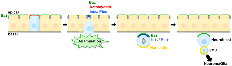
    1. An Y, Xue G, Shaobo Y, Mingxi D, Zhou X, Yu W, Ishibashi T, Zhang L, and Yan Y (2017). Apical constriction is driven by a pulsatile apical myosin network in delaminating Drosophila neuroblasts. Development 144, 2153–2164. - PubMed
    1. Baker NE (2017). Mechanisms of cell competition emerging from Drosophila studies. Curr. Opin. Cell Biol. 48, 40–46. - PMC - PubMed

Publication types

LinkOut - more resources