Removal of matrix-bound zoledronate prevents post-extraction osteonecrosis of the jaw by rescuing osteoclast function
- PMID: 29408511
- PMCID: PMC5878730
- DOI: 10.1016/j.bone.2018.01.030
Removal of matrix-bound zoledronate prevents post-extraction osteonecrosis of the jaw by rescuing osteoclast function
Abstract
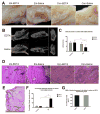
Unlike other antiresorptive medications, bisphosphonate molecules accumulate in the bone matrix. Previous studies of side-effects of anti-resorptive treatment focused mainly on systemic effects. We hypothesize that matrix-bound bisphosphonate molecules contribute to the pathogenesis of bisphosphonate-related osteonecrosis of the jaw (BRONJ). In this study, we examined the effect of matrix-bound bisphosphonates on osteoclast differentiation in vitro using TRAP staining and resorption assay, with and without pretreatment with EDTA. We also tested the effect of zoledronate chelation on the healing of post-extraction defect in rats. Our results confirmed that bisphosphonates bind to, and can be chelated from, mineralized matrix in vitro in a dose-dependent manner. Matrix-bound bisphosphonates impaired the differentiation of osteoclasts, evidenced by TRAP activity and resorption assay. Zoledronate-treated rats that underwent bilateral dental extraction with unilateral EDTA treatment showed significant improvement in mucosal healing and micro-CT analysis on the chelated sides. The results suggest that matrix-bound bisphosphonates are accessible to osteoclasts and chelating agents and contribute to the pathogenesis of BRONJ. The use of topical chelating agents is a promising strategy for the prevention of BRONJ following dental procedures in bisphosphonate-treated patients.
Keywords: Alveolar bone; Bisphosphonates; Bone matrix; Chelating agents; Osteonecrosis; Osteoporosis treatment; Zoledronate.
Copyright © 2018 Elsevier Inc. All rights reserved.
Figures


Similar articles
-
A Model for Osteonecrosis of the Jaw with Zoledronate Treatment following Repeated Major Trauma.PLoS One. 2015 Jul 17;10(7):e0132520. doi: 10.1371/journal.pone.0132520. eCollection 2015. PLoS One. 2015. PMID: 26186665 Free PMC article.
-
Teriparatide and the treatment of bisphosphonate-related osteonecrosis of the jaw: a rat model.Dentomaxillofac Radiol. 2014;43(1):20130144. doi: 10.1259/dmfr.20130144. Epub 2013 Oct 29. Dentomaxillofac Radiol. 2014. PMID: 24170800 Free PMC article.
-
Preexisting Periapical Inflammatory Condition Exacerbates Tooth Extraction-induced Bisphosphonate-related Osteonecrosis of the Jaw Lesions in Mice.J Endod. 2016 Nov;42(11):1641-1646. doi: 10.1016/j.joen.2016.07.020. Epub 2016 Sep 13. J Endod. 2016. PMID: 27637460 Free PMC article.
-
Clinical impact of bisphosphonates in root canal therapy.Saudi Med J. 2018 Mar;39(3):232-238. doi: 10.15537/smj.2018.3.20923. Saudi Med J. 2018. PMID: 29543299 Free PMC article. Review.
-
The role of bisphosphonates in medical oncology and their association with jaw bone necrosis.Oral Maxillofac Surg Clin North Am. 2014 May;26(2):231-7. doi: 10.1016/j.coms.2014.01.009. Oral Maxillofac Surg Clin North Am. 2014. PMID: 24794267 Review.
Cited by
-
Serum C-terminal cross-linking telopeptide level as a predictive biomarker of osteonecrosis after dentoalveolar surgery in patients receiving bisphosphonate therapy: Systematic review and meta-analysis.J Am Dent Assoc. 2019 Aug;150(8):664-675.e8. doi: 10.1016/j.adaj.2019.03.006. Epub 2019 Jun 28. J Am Dent Assoc. 2019. PMID: 31256803 Free PMC article.
-
Stress response in periodontal ligament stem cells may contribute to bisphosphonate‑associated osteonecrosis of the jaw: A gene expression array analysis.Mol Med Rep. 2020 Sep;22(3):2043-2051. doi: 10.3892/mmr.2020.11276. Epub 2020 Jun 26. Mol Med Rep. 2020. PMID: 32705175 Free PMC article.
-
Dendritic cell derived exosomes loaded with immunoregulatory cargo reprogram local immune responses and inhibit degenerative bone disease in vivo.J Extracell Vesicles. 2020 Aug 7;9(1):1795362. doi: 10.1080/20013078.2020.1795362. J Extracell Vesicles. 2020. PMID: 32944183 Free PMC article.
-
Dietary nitrate improves jaw bone remodelling in zoledronate-treated mice.Cell Prolif. 2023 Jul;56(7):e13395. doi: 10.1111/cpr.13395. Epub 2023 Feb 21. Cell Prolif. 2023. PMID: 36810909 Free PMC article.
-
Preclinical models of medication-related osteonecrosis of the jaw (MRONJ).Bone. 2021 Dec;153:116184. doi: 10.1016/j.bone.2021.116184. Epub 2021 Sep 11. Bone. 2021. PMID: 34520898 Free PMC article. Review.
References
-
- Russell RG. Bisphosphonates: the first 40 years. Bone. 2011;49(1):2–19. - PubMed
-
- Zarogoulidis K, Boutsikou E, Zarogoulidis P, Eleftheriadou E, Kontakiotis T, Lithoxopoulou H, et al. The impact of zoledronic acid therapy in survival of lung cancer patients with bone metastasis. Int J Cancer. 2009;125(7):1705–9. - PubMed
-
- Early Breast Cancer Trialists’ Collaborative G. Adjuvant bisphosphonate treatment in early breast cancer: meta-analyses of individual patient data from randomised trials. The Lancet. 386(10001):1353–61. - PubMed
-
- Oades GM, Coxon J, Colston KW. The potential role of bisphosphonates in prostate cancer. Prostate cancer and prostatic diseases. 2002;5(4):264–72. - PubMed
Publication types
MeSH terms
Substances
Grants and funding
LinkOut - more resources
Full Text Sources
Other Literature Sources

