. 2018 May 15:172:437-449.
doi: 10.1016/j.neuroimage.2018.01.082. Epub 2018 Feb 2.

Triple representation of language, working memory, social and emotion processing in the cerebellum: convergent evidence from task and seed-based resting-state fMRI analyses in a single large cohort

Affiliations

Triple representation of language, working memory, social and emotion processing in the cerebellum: convergent evidence from task and seed-based resting-state fMRI analyses in a single large cohort

Xavier Guell et al. Neuroimage. .

Abstract

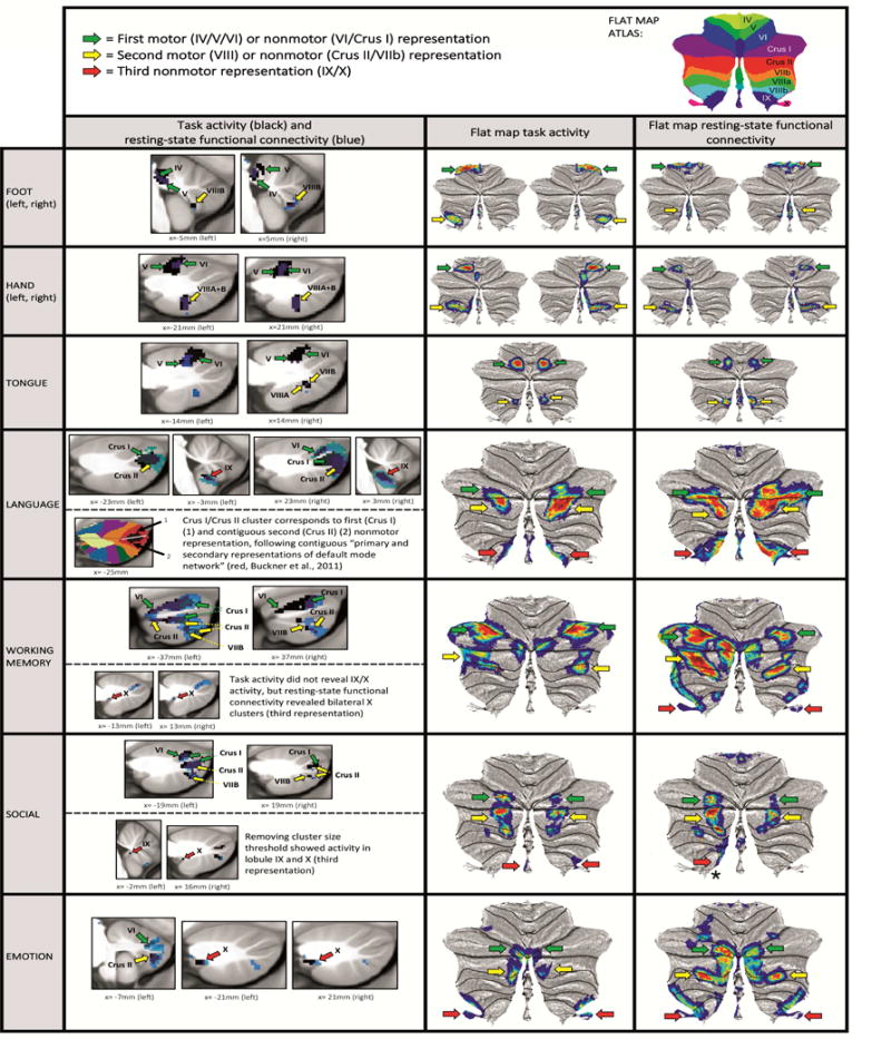
Delineation of functional topography is critical to the evolving understanding of the cerebellum's role in a wide range of nervous system functions. We used data from the Human Connectome Project (n = 787) to analyze cerebellar fMRI task activation (motor, working memory, language, social and emotion processing) and resting-state functional connectivity calculated from cerebral cortical seeds corresponding to the peak Cohen's d of each task contrast. The combination of exceptional statistical power, activation from both motor and multiple non-motor tasks in the same participants, and convergent resting-state networks in the same participants revealed novel aspects of the functional topography of the human cerebellum. Consistent with prior studies there were two distinct representations of motor activation. Newly revealed were three distinct representations each for working memory, language, social, and emotional task processing that were largely separate for these four cognitive and affective domains. In most cases, the task-based activations and the corresponding resting-network correlations were congruent in identifying the two motor representations and the three non-motor representations that were unique to working memory, language, social cognition, and emotion. The definitive localization and characterization of distinct triple representations for cognition and emotion task processing in the cerebellum opens up new basic science questions as to why there are triple representations (what different functions are enabled by the different representations?) and new clinical questions (what are the differing consequences of lesions to the different representations?).

Keywords: Cerebellar topography; Cognition; Resting state fMRI; Sensorimotor; Task-based fMRI.


Figures

Fig.1
Fig.1
Convergence of findings from multiple studies of cerebellar topography suggesting an overarching organizing principle based on two motor and three nonmotor representations. Green circles indicate first motor (lobules IV/V/VI) and nonmotor (VI/Crus I) representation; blue circles indicate second motor (lobule VIII) and nonmotor (Crus II/VIIB) representation; red circles indicate third nonmotor representation (lobules IX/X). Note that areas of first and second nonmotor representation are contiguous. A: Classical electrical stimulation studies showed double representation of sensorimotor activation in the cerebellum (first = lobules IV/V/VI and second = lobule VIII) (Snider and Eldred, 1952; permission pending). B: Tract tracing studies demonstrated labeling of the cerebellum in two different locations after injecting viral tracers in motor and nonmotor cerebral cortical areas (viral tracer in M1 labeled cerebellar lobules IV/V/VI and VIIB/VIII, left image; viral tracer in prefrontal cortex area 46 labeled cerebellar lobules Crus I / Crus II and IX, right image) (Kelly and Strick, 2003; permission pending). C: Resting-state functional connectivity studies suggest that each resting-state network is represented three times in the cerebellum (approximately lobules IV/V/VI/Crus I, lobules Crus II/VIIB/VIII and lobules IX/X) with the possible exception of the somatomotor network (represented only twice) (image from Buckner et al., 2011, where each color represents one of the seven resting-state networks defined in Yeo et al., 2011).
Fig. 2
Fig. 2
Summary of cerebellar activation for motor (red), working memory (blue), language (yellow), social processing (green) and emotion processing (magenta); coronal plane. (H) = hand, (F) = foot, (T) = tongue. Activation maps are thresholded at a voxel-level threshold of d>0.5. Only clusters >100 mmˆ3 are shown. Left is shown on the left.
Fig. 3
Fig. 3
Grey dots mark the seed from which resting-state functional connectivity was calculated for each task contrast. Working memory seed Cohen’s d=1.37. Language d=1.34. Emotion d=1.33. Right hand d=2.75. Left hand d=2.87. Right foot d=2.54. Left foot d=2.62. Tongue d=3.08. Regions shown in black (emotion) and white (social) had a higher task contrast Cohen’s d value than the selected seed location, but resting-state functional connectivity calculated from those seeds did not show overlap with areas of task activation in the cerebellum.
Fig. 4
Fig. 4
Motor task contrasts revealed double representation (first = lobules IV/V/VI and second = lobule VIII); and nonmotor task contrasts revealed triple representation (first = lobules VI/Crus I; second = lobules Crus II/VIIB; and third = lobules IX/X), except for working memory task activation, which did not reveal a third representation. Resting-state functional connectivity overlapped with clusters of task activation, also revealing a pattern of double motor and triple nonmotor representation. First and second nonmotor representations are sometimes separate (e.g. working memory) and sometimes contiguous (e.g. language processing). Key. First column images: Black = cerebellar task activation (thresholded at d>0.5 [medium effect size] and cluster size>100mmˆ3). Blue = resting-state functional connectivity calculated from cerebral cortical seed for each task contrast (thresholded at Fisher’s z>0.309, equivalent to r>0.3 [medium effect size]). Second and third column images (flat maps) represent the same resting-state functional connectivity and task activation clusters with no cluster size threshold. Note that cluster size threshold removal does not violate our statistical approach (see section 2.4). Green arrows correspond to first motor or nonmotor representation, yellow arrows correspond to second motor or nonmotor representation, red arrows correspond to third nonmotor representation. Red arrow with an asterisk in social processing resting-state connectivity indicates lobule IX/X engagement that does not overlap with social processing task activation.

References

    1. Adamaszek M, D’Agata F, Ferrucci R, et al. Consensus Paper: Cerebellum and Emotion. Cerebellum. 2017;16(2):552–576. - PubMed
    1. Baillieux H, De Smet HJ, Paquier PF, De Deyn PP, Mariën P. Cerebellar neurocognition: insights into the bottom of the brain. Clin Neurol Neurosurg. 2008;110(8):763–73. - PubMed
    1. Barch DM, Burgess GC, Harms MP, et al. Function in the human connectome: task-fMRI and individual differences in behavior. Neuroimage. 2013;80:169–89. - PMC - PubMed
    1. Baumann O, Mattingley JB. Functional topography of primary emotion processing in the human cerebellum. Neuroimage. 2012;61(4):805–11. - PubMed
    1. Baumann O, Borra RJ, Bower JM, et al. Consensus paper: the role of the cerebellum in perceptual processes. Cerebellum. 2015;14(2):197–220. - PMC - PubMed

Publication types