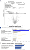
Fetal regional brain protein signature in FASD rat model
- PMID: 29408587
- PMCID: PMC5834402
- DOI: 10.1016/j.reprotox.2018.01.004
Fetal regional brain protein signature in FASD rat model
Abstract
Fetal alcohol spectrum disorders (FASD) describe neurodevelopmental deficits in children exposed to alcohol in utero. We hypothesized that gestational alcohol significantly alters fetal brain regional protein signature. Pregnant rats were binge-treated with alcohol or pair-fed and nutritionally-controlled. Mass spectrometry identified 1806, 2077, and 1456 quantifiable proteins in the fetal hippocampus, cortex, and cerebellum, respectively. A stronger effect of alcohol exposure on the hippocampal proteome was noted: over 600 hippocampal proteins were significantly (P < .05) altered, including annexin A2, nucleobindin-1, and glypican-4, regulators of cellular growth and developmental morphogenesis. In the cerebellum, cadherin-13, reticulocalbin-2, and ankyrin-2 (axonal growth regulators) were significantly (P < .05) altered; altered cortical proteins were involved in autophagy (endophilin-B1, synaptotagmin-1). Ingenuity analysis identified proteins involved in protein homeostasis, oxidative stress, mitochondrial dysfunction, and mTOR as major pathways in the cortex and hippocampus significantly (P < .05) affected by alcohol. Thus, neurodevelopmental protein changes may directly relate to FASD neuropathology.
Keywords: Alcohol; FASD; Fetal; Teratology.
Copyright © 2018 Elsevier Inc. All rights reserved.
Figures


Similar articles
-
Intrauterine environment-genome interaction and children's development (1): Ethanol: a teratogen in developing brain.J Toxicol Sci. 2009;34 Suppl 2:SP273-8. doi: 10.2131/jts.34.sp273. J Toxicol Sci. 2009. PMID: 19571480 Review.
-
Placental Proteomics Reveal Insights into Fetal Alcohol Spectrum Disorders.Alcohol Clin Exp Res. 2017 Sep;41(9):1551-1558. doi: 10.1111/acer.13448. Epub 2017 Aug 9. Alcohol Clin Exp Res. 2017. PMID: 28722160 Free PMC article.
-
Molecular changes during neurodevelopment following second-trimester binge ethanol exposure in a mouse model of fetal alcohol spectrum disorder: from immediate effects to long-term adaptation.Dev Neurosci. 2014;36(1):29-43. doi: 10.1159/000357496. Epub 2014 Jan 24. Dev Neurosci. 2014. PMID: 24481079
-
Hippocampal transcriptome reveals novel targets of FASD pathogenesis.Brain Behav. 2019 Jul;9(7):e01334. doi: 10.1002/brb3.1334. Epub 2019 May 29. Brain Behav. 2019. PMID: 31140755 Free PMC article.
-
Potential roles of imprinted genes in the teratogenic effects of alcohol on the placenta, somatic growth, and the developing brain.Exp Neurol. 2022 Jan;347:113919. doi: 10.1016/j.expneurol.2021.113919. Epub 2021 Nov 6. Exp Neurol. 2022. PMID: 34752786 Review.
Cited by
-
Dose-related shifts in proteome and function of extracellular vesicles secreted by fetal neural stem cells following chronic alcohol exposure.Heliyon. 2022 Nov 1;8(11):e11348. doi: 10.1016/j.heliyon.2022.e11348. eCollection 2022 Nov. Heliyon. 2022. PMID: 36387439 Free PMC article.
-
The Impact of Oxidative Stress on Pediatrics Syndromes.Antioxidants (Basel). 2022 Oct 5;11(10):1983. doi: 10.3390/antiox11101983. Antioxidants (Basel). 2022. PMID: 36290706 Free PMC article. Review.
-
Mechanisms of Alcohol Interference with Hippocampal Neurogenesis and its Repair Strategies.Mol Neurobiol. 2025 Jul 17. doi: 10.1007/s12035-025-05240-6. Online ahead of print. Mol Neurobiol. 2025. PMID: 40676366 Review.
-
Aversive Learning Deficits and Depressive-Like Behaviors Are Accompanied by an Increase in Oxidative Stress in a Rat Model of Fetal Alcohol Spectrum Disorders: The Protective Effect of Rapamycin.Int J Mol Sci. 2021 Jun 30;22(13):7083. doi: 10.3390/ijms22137083. Int J Mol Sci. 2021. PMID: 34209274 Free PMC article.
-
Interaction of alcohol & phosphatidic acid in maternal rat uterine artery function.Reprod Toxicol. 2022 Aug;111:178-183. doi: 10.1016/j.reprotox.2022.05.017. Epub 2022 Jun 6. Reprod Toxicol. 2022. PMID: 35671880 Free PMC article.
References
-
- Sokol RJ, Delaney-Black V, Nordstrom B. Fetal alcohol spectrum disorder. JAMA. 2003;290:2996–2999. - PubMed
-
- Roozen S, et al. Worldwide Prevalence of Fetal Alcohol Spectrum Disorders: A Systematic Literature Review Including Meta-Analysis. Alcohol Clin Exp Res. 2016;40:18–32. - PubMed
-
- Tan CH, Denny CH, Cheal NE, Sniezek JE, Kanny D. Alcohol use and binge drinking among women of childbearing age – United States, 2011–2013. MMWR Morb Mortal Wkly Rep. 2015;64:1042–1046. - PubMed
Publication types
MeSH terms
Substances
Grants and funding
LinkOut - more resources
Full Text Sources
Other Literature Sources
Medical
Miscellaneous

