. 2018 May;154(6):1751-1763.e2.
doi: 10.1053/j.gastro.2018.01.042. Epub 2018 Feb 1.

Microbiota-Derived Metabolic Factors Reduce Campylobacteriosis in Mice

Affiliations

Microbiota-Derived Metabolic Factors Reduce Campylobacteriosis in Mice

Xiaolun Sun et al. Gastroenterology. 2018 May.

Abstract

Background & aims: Campylobacter jejuni, a prevalent foodborne bacterial pathogen, exploits the host innate response to induce colitis. Little is known about the roles of microbiota in C jejuni-induced intestinal inflammation. We investigated interactions between microbiota and intestinal cells during C jejuni infection of mice.

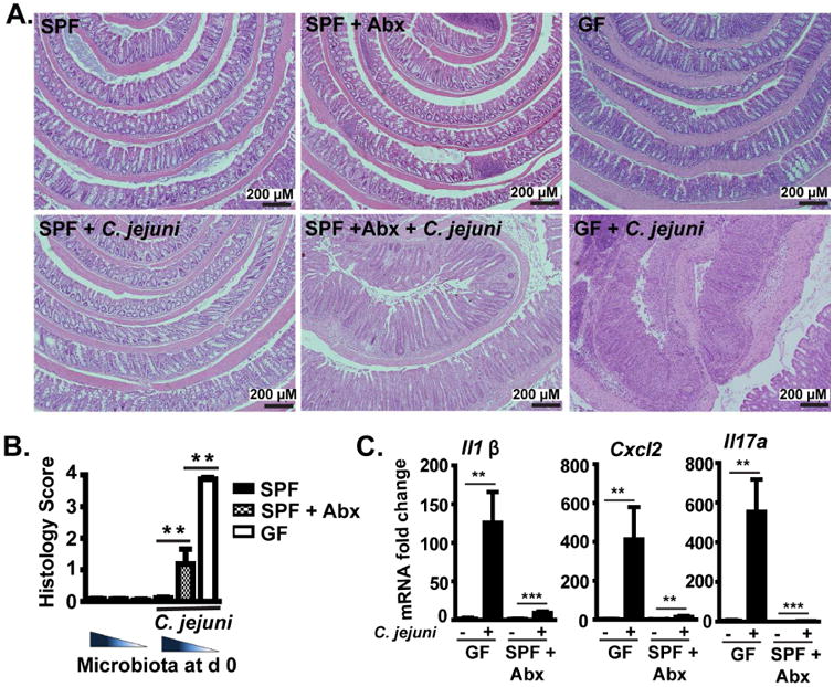
Methods: Germ-free C57BL/6 Il10-/- mice were colonized with conventional microbiota and infected with a single dose of C jejuni (109 colony-forming units/mouse) via gavage. Conventional microbiota were cultured under aerobic, microaerobic, or anaerobic conditions and orally transplanted into germ-free Il10-/- mice. Colon tissues were collected from mice and analyzed by histology, real-time polymerase chain reaction, and immunoblotting. Fecal microbiota and bile acids were analyzed with 16S sequencing and high-performance liquid chromatography with mass spectrometry, respectively.

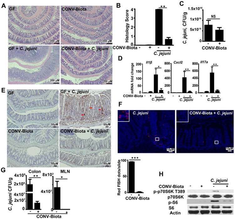
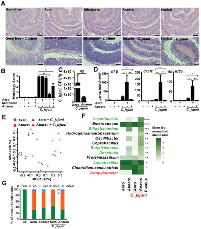
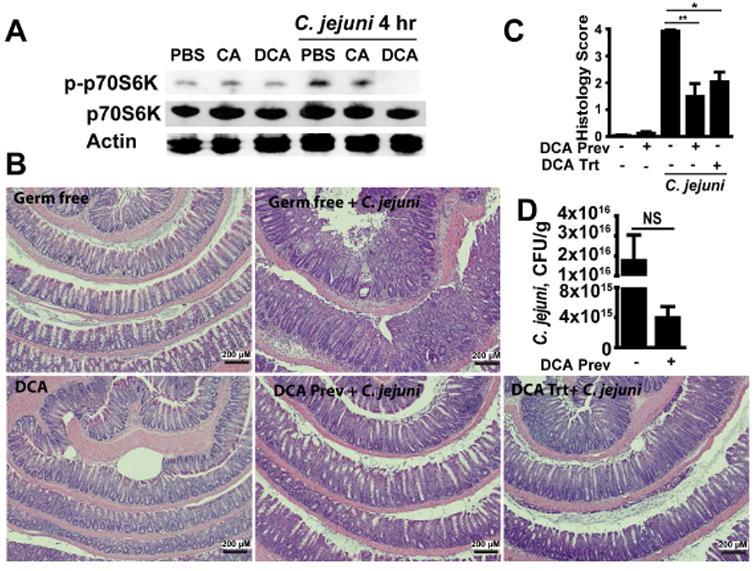
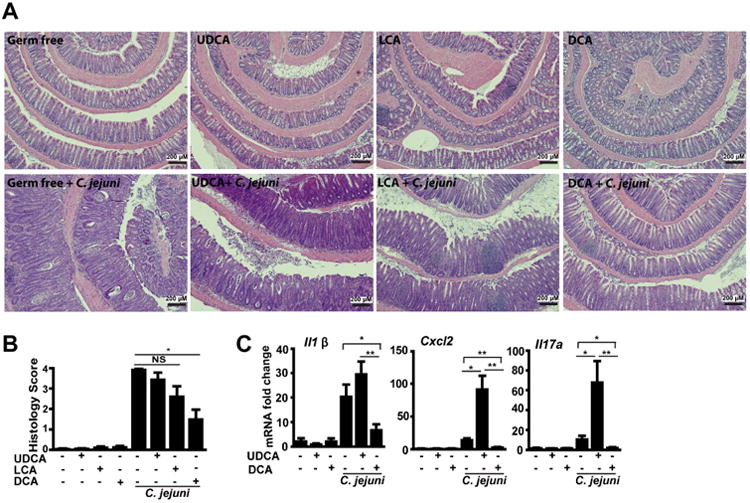
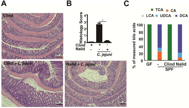
Results: Introduction of conventional microbiota reduced C jejuni-induced colitis in previously germ-free Il10-/- mice, independent of fecal load of C jejuni, accompanied by reduced activation of mammalian target of rapamycin. Microbiota transplantation and 16S ribosomal DNA sequencing experiments showed that Clostridium XI, Bifidobacterium, and Lactobacillus were enriched in fecal samples from mice colonized with microbiota cultured in anaerobic conditions (which reduce colitis) compared with mice fed microbiota cultured under aerobic conditions (susceptible to colitis). Oral administration to mice of microbiota-derived secondary bile acid sodium deoxycholate, but not ursodeoxycholic acid or lithocholic acid, reduced C jejuni-induced colitis. Depletion of secondary bile acid-producing bacteria with antibiotics that kill anaerobic bacteria (clindamycin) promoted C jejuni-induced colitis in specific pathogen-free Il10-/- mice compared with the nonspecific antibiotic nalidixic acid; colitis induction by antibiotics was associated with reduced level of luminal deoxycholate.

Conclusions: We identified a mechanism by which the microbiota controls susceptibility to C jejuni infection in mice, via bacteria-derived secondary bile acids.

Keywords: DCA; Infection; Metabolism; Microbiome.


Conflict of interest statement

Conflict of interest: The authors declare no conflict of interest.

Figures

Figure 1
Figure 1. Pan-depletion of microbiota exacerbates C. jejuni-induced intestinal inflammation in Il10-/- mice
Cohorts of 5-9 GF or SPF Il10-/- mice were gavaged with a single dose of 109 CFU C. jejuni/mouse and were euthanized 12 or 21 days post-infection. SPF Il10-/-mice were treated with an antibiotics (Abx) cocktail in their drinking water for 7 days before infection. (A) H&E staining showing representative intestinal histology of C. jejuni-induced colitis in SPF (left), SPF+ Abx (middle) and GF (GF, right) Il10-/- mice. (B) Quantification of histological intestinal damage score. (C) Colonic Il1β, Cxcl2, and Il17a mRNA qPCR fold change relative to GF and normalized to Gapdh. **, P<0.01. Scale bar is 200 μm. Results are representative of 3 independent experiments.
Figure 2
Figure 2. Microbiota prevents C. jejuni-induced intestinal inflammation in GF Il10-/- mice in a manner independent of luminal colonization exclusion
Cohorts of 5-6 GF Il10-/- mice were colonized with a conventionalized microbiota (CONV-Biota) for 14 days or left in GF conditions. The mice were then infected with a single dose of 109 CFU C. jejuni/mouse and were euthanized 12 days post-infection. (A) Representative intestinal histology images. (B) Quantification of histological intestinal damage score. (C) C. jejuni colonic luminal colonization level using culture. (D) Colonic Il1β, Cxcl2, and Il17a mRNA qPCR fold change relative to GF and normalized to Gapdh. (E) Immunohistochemistry of myeloperoxidase positive neutrophils (brown dots). Red arrows indicate neutrophil accumulation in the crypt lumen and formation of crypt abscesses. (F) Presence of C. jejuni (red dots, counted as dots/slide (lower panel)) in colonic sections of infected mice, detected using fluorescence in situ hybridization (FISH) assay. (G) Live C. jejuni count in the colon tissue (left panel) and MLN (right panel). Briefly, MLN and colon tissue were aseptically resected, weighed, homogenized in PBS, serially diluted, and plated on Campylobacter-selective blood plates. The bacteria were counted after 48 h at 37°C using the GasPak system. (H) Western blot of total and phosphorylated p70S6 (T389) and S-6 (S235/236) and Actin from resected colon tissues. Scale bar is 200 (Fig. A), 50 (Fig. E) or 10 (Fig. F) μm. All graphs depict mean ± SEM. NS, not significant, *, P<0.05; **, P<0.01. Results are representative of 3 independent experiments.
Figure 3
Figure 3. Anaerobic bacteria are enriched in campylobacteriosis resistant mice
Stool samples from mice conventionalized for 3 or 14 days and then infected with C. jejuni were subjected to 16S rDNA sequencing 12 days after C. jejuni infection (see Supplementary Figure 3). (A) PCoA comparing the microbiome composition of C. jejuni infected mice conventionalized for 3 or 14 days. Histological inflammation scores are shown as color code. PCoA 1 FDR-P = 0.01 (linear mixed effect model) and 1.26e-05 (t-test) and cage FDR-P= 0.97 (linear mixed effect) (B) Shannon diversity and (C) Choa1 richness show no differences or correlation with histological inflammation scores (FDR-P > 0.05 linear mixed effect and t-test). Histological inflammation scores are shown as color code (D) Heatmap representation of genera significantly different (FDR-P < 0.05, t-test) between 3 days conventionalized and 14 days conventionalized Il10-/- mice prior to C. jejuni infection. Majority of the genera enriched in campylobacteriosis resistant mice are anaerobic. Fac anaerobic: facultative anaerobic.
Figure 4
Figure 4. Anaerobic microbiota isolated from CONV-Biota attenuates C. jejuni-induced colitis
Cohorts of 4-8 GF Il10-/- mice were colonized with microbiota cultured under aerobic (Aero), microaerobic (Microaero), or anaerobic (Anaero) conditions, or all three groups pooled for 14 days. The mice were then gavaged with a single dose of 109 CFU C. jejuni/mouse and were euthanized 12 days post-infection. Stool samples from Anaero- and Aero-Biota mice were subjected to 16S rDNA sequencing and HPLC/MS analysis of bile acids. (A) H&E staining showing representative intestinal histology of C. jejuni-induced colitis in Il10-/- mice. (B) Quantification of histological intestinal damage score. (C) C. jejuni colonic luminal colonization level using culture in mice colonized with Aero- or Anaero-Biota. (D) Colonic Il1β, Cxcl2, and Il17a mRNA qPCR fold change relative to GF and normalized to Gapdh. (E) PCoA comparing Anaero- and Aero-Biota microbiome composition pre- and post-C. jejuni infection, based on 16S rDNA sequencing. (F) Heatmap representation of genera significantly different between Anaero-and Aero-Biota-colonized Il10-/- mice following C. jejuni infection, plus Campylobacter (red), which was not significant. Their abundance prior to infection is also shown. Green font indicates genera associated with anti-inflammatory response. Asterisks indicate the p-value after multiple hypothesis correction. (G) Relative stool bile acid profile measured by HPLC/MS. TCA, taurocholic acid and tauromuricholic acid; CA, cholate; LCA, lithocholic acid; UDCA, Ursodeoxycholic acid; DCA, deoxycholate. ****, P < 0.0001; ***, P < 0.001; **, P < 0.01; *, P < 0.05; NS, not significant. Scale bar is 200 μm. Results are representative of 3 independent experiments.
Figure 5
Figure 5. The microbial metabolite deoxycholate inhibits mTOR activity and prevents and treats C. jejuni-induced colitis
(A) Splenocytes isolated from Il10-/- mice were infected with C. jejuni (multiplicity of infection 50) and cultured in the presence of CA and DCA. Total and phosphorylated p70S6K (T389) and Actin was measured by Western Blot. (B) Cohorts of 4-7 GF Il10-/- mice infected with a single dose of 109 CFU C. jejuni/mouse were gavaged with the secondary bile acid DCA daily (DCA prevention (Prev)). For DCA treatment (DCA trt), mice infected with C. jejuni were gavaged with DCA on days 5-12 post-infection. H&E staining showing representative day 12 intestinal histology of C. jejuni-induced colitis in Il10-/- mice. (C) Quantification of histological intestinal damage score. (D) C. jejuni colonic luminal colonization level using culture. **, P < 0.01; *, P < 0.05; NS, not significant. Scale bar is 200 μm. Results are representative of 3 independent experiments.
Figure 6
Figure 6. Microbial metabolites of the secondary bile acids LCA and UDCA minimally impact C. jejuni-induced colitis
Cohorts of 4-7 GF Il10-/- mice infected with a single dose of 109 CFU C. jejuni/mouse were gavaged with the secondary bile acids UDCA, LCA or DCA daily. The mice were then euthanized 12-days after infection. (A) H&E staining showing representative intestinal histology of C. jejuni-induced colitis in Il10-/- mice. (B) Quantification of histological intestinal damage score. (C) Colonic Il1β, Cxcl2, and Il17a mRNA qPCR fold change relative to GF and normalized to Gapdh. **, P<0.01; *, P<0.05; NS, not significant. Scale bar is 200 μm. Results are representative of 3 independent experiments.
Figure 7
Figure 7. Targeted depletion of secondary bile acid-metabolizing microbiota promotes Il10-/- mouse susceptibility to C. jejuni
Cohorts of 4 SPF Il10-/- mice gavaged with the antibiotics clindamycin (Clind) or nalidixic acid (Nalid) for 7 days prior to infection with a single dose of 109 CFU C. jejuni/mouse. (A) H&E staining showing representative intestinal histology of C. jejuni-induced colitis in Il10-/- mice 21 days post-infection. (B) Quantification of histological intestinal damage score. (C) Relative stool bile acid profile measured by HPLC/MS (see Supplementary Table 4 for values). *, P<0.05. Scale bar is 200 μm. Results are representative of 3 independent experiments.

References

    1. Kaakoush NO, Castano-Rodriguez N, Mitchell HM, et al. Global Epidemiology of Campylobacter Infection. Clin Microbiol Rev. 2015;28:687–720. - PMC - PubMed
    1. Blaser MJ. Epidemiologic and clinical features of Campylobacter jejuni infections. J Infect Dis. 1997;176(Suppl 2):S103–5. - PubMed
    1. van Spreeuwel JP, Duursma GC, Meijer CJ, et al. Campylobacter colitis: histological immunohistochemical and ultrastructural findings. Gut. 1985;26:945–51. - PMC - PubMed
    1. Lippert E, Karrasch T, Sun X, et al. Gnotobiotic IL-10; NF-kappaB mice develop rapid and severe colitis following Campylobacter jejuni infection. PLoS One. 2009;4:e7413. - PMC - PubMed
    1. Sun X, Liu B, Sartor RB, et al. Phosphatidylinositol 3-kinase-gamma signaling promotes Campylobacter jejuni-induced colitis through neutrophil recruitment in mice. J Immunol. 2013;190:357–65. - PMC - PubMed

Publication types

MeSH terms