

Quantitative Primary Tumor Indocyanine Green Measurements Predict Osteosarcoma Metastatic Lung Burden in a Mouse Model
- PMID: 29408832
- PMCID: PMC6260021
- DOI: 10.1007/s11999.0000000000000003
Quantitative Primary Tumor Indocyanine Green Measurements Predict Osteosarcoma Metastatic Lung Burden in a Mouse Model
Abstract
Background: Current preclinical osteosarcoma (OS) models largely focus on quantifying primary tumor burden. However, most fatalities from OS are caused by metastatic disease. The quantification of metastatic OS currently relies on CT, which is limited by motion artifact, requires intravenous contrast, and can be technically demanding in the preclinical setting. We describe the ability for indocyanine green (ICG) fluorescence angiography to quantify primary and metastatic OS in a previously validated orthotopic, immunocompetent mouse model.
Questions/purposes: (1) Can near-infrared ICG fluorescence be used to attach a comparable, quantitative value to the primary OS tumor in our experimental mouse model? (2) Will primary tumor fluorescence differ in mice that go on to develop metastatic lung disease? (3) Does primary tumor fluorescence correlate with tumor volume measured with CT?
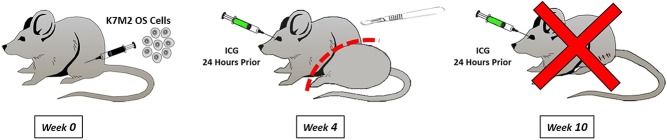
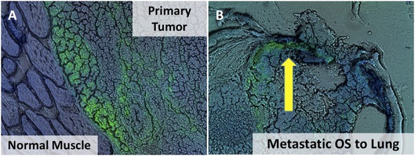
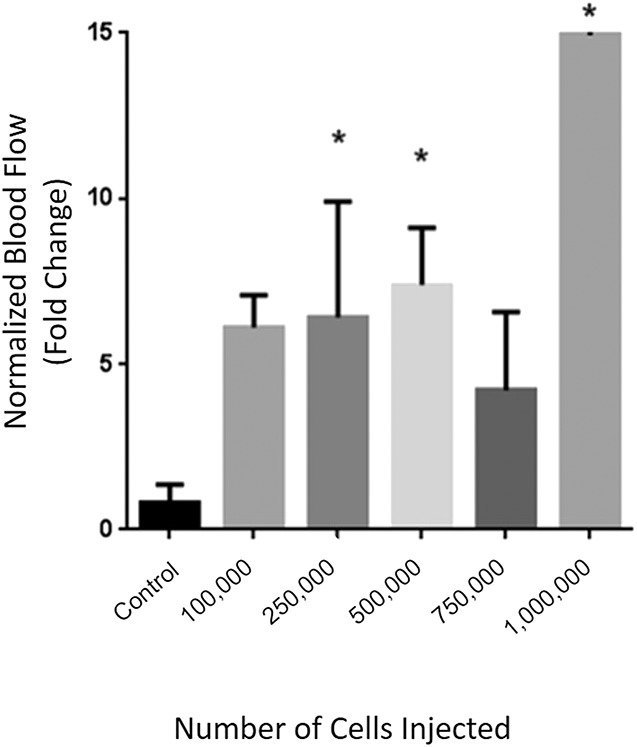
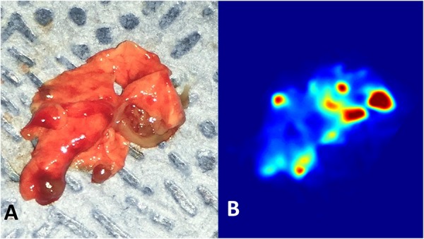
Methods: Six groups of 4- to 6-week-old immunocompetent Balb/c mice (n = 6 per group) received paraphyseal injections into their left hindlimb proximal tibia consisting of variable numbers of K7M2 mouse OS cells. A hindlimb transfemoral amputation was performed 4 weeks after injection with euthanasia and lung extraction performed 10 weeks after injection. Histologic examination of lung and primary tumor specimens confirmed ICG localization only within the tumor bed.
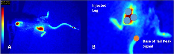
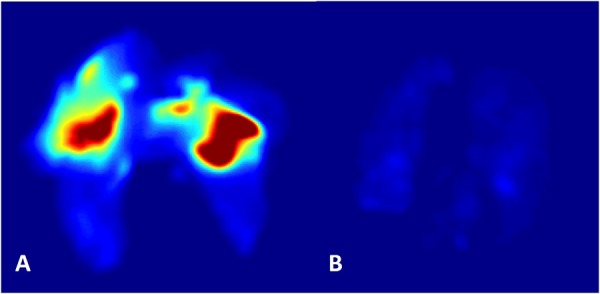
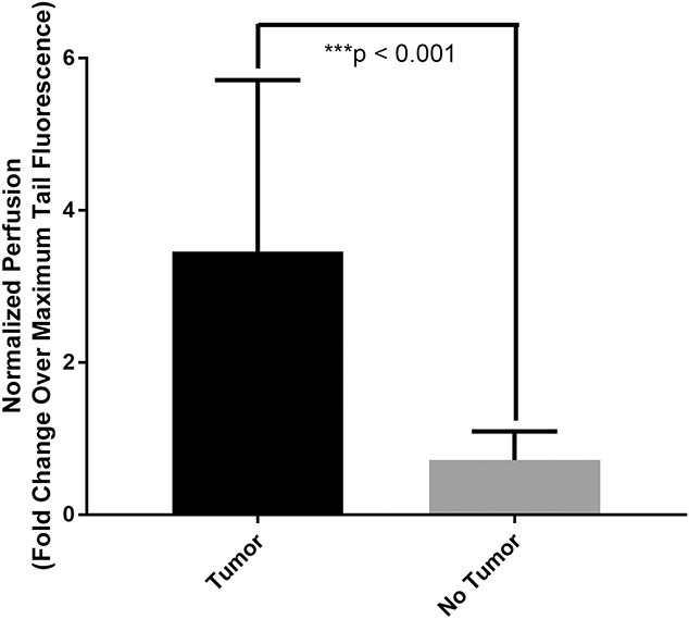
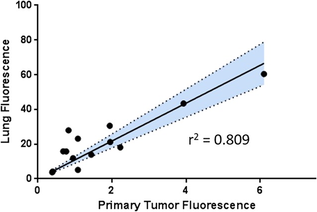
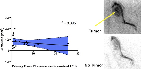
Results: Mice with visible or palpable tumor growth had greater hindlimb fluorescence (3.5 ± 2.3 arbitrary perfusion units [APU], defined as the fluorescence pixel return normalized by the detector) compared with those with a negative examination (0.71 ± 0.38 APU, -2.7 ± 0.5 mean difference, 95% confidence interval -3.7 to -1.8, p < 0.001). A strong linear trend (r = 0.81, p < 0.01) was observed between primary tumor and lung fluorescence, suggesting that quantitative ICG tumor fluorescence is directly related to eventual metastatic burden. We did not find a correlation (r = 0.04, p = 0.45) between normalized primary tumor fluorescence and CT volumetric measurements.
Conclusions: We demonstrate a novel methodology for quantifying primary and metastatic OS in a previously validated, immunocompetent, orthotopic mouse model. Quantitative fluorescence of the primary tumor with ICG angiography is linearly related to metastatic burden, a relationship that does not exist with respect to clinical tumor size. This highlights the potential utility of ICG near-infrared fluorescence imaging as a valuable preclinical proof-of-concept modality. Future experimental work will use this model to evaluate the efficacy of novel OS small molecule inhibitors.
Clinical relevance: Given the histologic localization of ICG to only the tumor bed, we envision the clinical use of ICG angiography as an intraoperative margin and tumor detector. Such a tool may be used as an alternative to intraoperative histology to confirm negative primary tumor margins or as a valuable tool for debulking surgeries in vulnerable anatomic locations. Because we have demonstrated the successful preservation of ICG in frozen tumor samples, future work will focus on blinded validation of this modality in observational human trials, comparing the ICG fluorescence of harvested tissue samples with fresh frozen pathology.
Conflict of interest statement
All ICMJE Conflict of Interest Forms for authors and
Figures
Comment in
-
CORR Insights®: Quantitative Primary Tumor Indocyanine Green Measurements Predict Osteosarcoma Metastatic Lung Burden in a Mouse Model.Clin Orthop Relat Res. 2018 Mar;476(3):488-489. doi: 10.1007/s11999.0000000000000136. Clin Orthop Relat Res. 2018. PMID: 29529629 Free PMC article. No abstract available.
Similar articles
-
Disulfiram reduces metastatic osteosarcoma tumor burden in an immunocompetent Balb/c or-thotopic mouse model.Oncotarget. 2018 Jul 10;9(53):30163-30172. doi: 10.18632/oncotarget.25733. eCollection 2018 Jul 10. Oncotarget. 2018. PMID: 30046395 Free PMC article.
-
Tumor Resection Guided by Intraoperative Indocyanine Green Dye Fluorescence Angiography Results in Negative Surgical Margins and Decreased Local Recurrence in an Orthotopic Mouse Model of Osteosarcoma.Ann Surg Oncol. 2019 Mar;26(3):894-898. doi: 10.1245/s10434-018-07114-9. Epub 2018 Dec 27. Ann Surg Oncol. 2019. PMID: 30588559 Free PMC article.
-
Intratibial Injection Causes Direct Pulmonary Seeding of Osteosarcoma Cells and Is Not a Spontaneous Model of Metastasis: A Mouse Osteosarcoma Model.Clin Orthop Relat Res. 2018 Jul;476(7):1514-1522. doi: 10.1007/s11999.0000000000000291. Clin Orthop Relat Res. 2018. PMID: 29601385 Free PMC article.
-
[A new approach for studying the retinal and choroidal circulation].Nippon Ganka Gakkai Zasshi. 2004 Dec;108(12):836-61; discussion 862. Nippon Ganka Gakkai Zasshi. 2004. PMID: 15656089 Review. Japanese.
-
Intraoperative Indocyanine Green (ICG) Angiography for the Identification of the Parathyroid Glands: Current Evidence and Future Perspectives.In Vivo. 2020 Jan-Feb;34(1):23-32. doi: 10.21873/invivo.11741. In Vivo. 2020. PMID: 31882459 Free PMC article. Review.
Cited by
-
Progresses in Fluorescence Imaging Guidance for Bone and Soft Tissue Sarcoma Surgery.Front Oncol. 2022 Jul 4;12:879697. doi: 10.3389/fonc.2022.879697. eCollection 2022. Front Oncol. 2022. PMID: 35860548 Free PMC article. Review.
-
CSCs: regenerating optimism for osteosarcoma treatment.Oncotarget. 2018 Aug 3;9(60):31562-31563. doi: 10.18632/oncotarget.25820. eCollection 2018 Aug 3. Oncotarget. 2018. PMID: 30167075 Free PMC article. No abstract available.
-
Disulfiram reduces metastatic osteosarcoma tumor burden in an immunocompetent Balb/c or-thotopic mouse model.Oncotarget. 2018 Jul 10;9(53):30163-30172. doi: 10.18632/oncotarget.25733. eCollection 2018 Jul 10. Oncotarget. 2018. PMID: 30046395 Free PMC article.
-
Tumor Resection Guided by Intraoperative Indocyanine Green Dye Fluorescence Angiography Results in Negative Surgical Margins and Decreased Local Recurrence in an Orthotopic Mouse Model of Osteosarcoma.Ann Surg Oncol. 2019 Mar;26(3):894-898. doi: 10.1245/s10434-018-07114-9. Epub 2018 Dec 27. Ann Surg Oncol. 2019. PMID: 30588559 Free PMC article.
-
A Novel Orthotopic Implantation Technique for Osteosarcoma Produces Spontaneous Metastases and Illustrates Dose-Dependent Efficacy of B7-H3-CAR T Cells.Front Immunol. 2021 Jun 15;12:691741. doi: 10.3389/fimmu.2021.691741. eCollection 2021. Front Immunol. 2021. PMID: 34211478 Free PMC article.
References
-
- Allen RC, Oetting TA. Indocyanine green toxicity. Ophthalmology. 2007;114:197; author reply 197. - PubMed
-
- Anderson ME. Update on survival in osteosarcoma. Orthop Clin North Am. 2016;47:283–292. - PubMed
-
- Bacci G, Longhi A, Versari M, Mercuri M, Briccoli A, Picci P. Prognostic factors for osteosarcoma of the extremity treated with neoadjuvant chemotherapy: 15-year experience in 789 patients treated at a single institution. Cancer. 2006;106:1154–1161. - PubMed
-
- Cole HA, Ichikawa J, Colvin DC, O'Rear L, Schoenecker JG. Quantifying intra-osseous growth of osteosarcoma in a murine model with radiographic analysis. J Orthop Res. 2011;29:1957–1962. - PubMed
-
- Fourman MS, Gersch RP, Levites HA, Phillips BT, Bui DT. Is there a right way to interpret SPY? Normalization of indocyanine green angiography readings in a burn model. Plast Reconstr Surg. 2015;136:128e-130e. - PubMed
MeSH terms
Substances
Grants and funding
LinkOut - more resources
Full Text Sources
Other Literature Sources
Medical
