. 2018 Feb 6;14(2):e1007178.
doi: 10.1371/journal.pgen.1007178. eCollection 2018 Feb.

Axin phosphorylation in both Wnt-off and Wnt-on states requires the tumor suppressor APC

Affiliations

Axin phosphorylation in both Wnt-off and Wnt-on states requires the tumor suppressor APC

Ofelia Tacchelly-Benites et al. PLoS Genet. .

Abstract

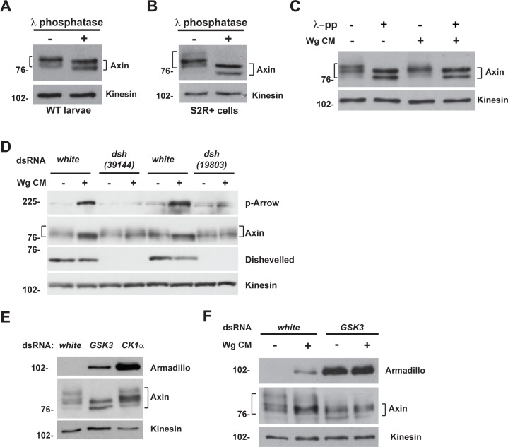
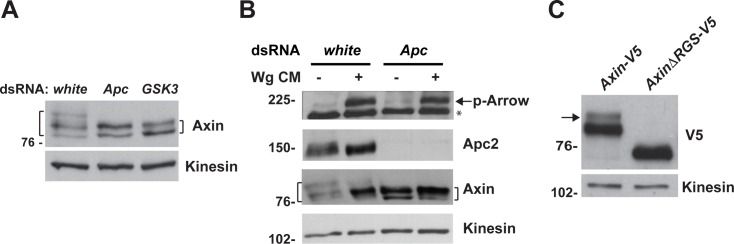
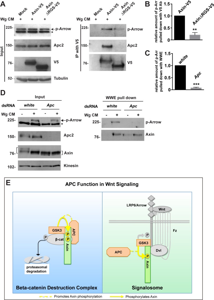
The aberrant activation of Wnt signal transduction initiates the development of 90% of colorectal cancers, the majority of which arise from inactivation of the tumor suppressor Adenomatous polyposis coli (APC). In the classical model for Wnt signaling, the primary role of APC is to act, together with the concentration-limiting scaffold protein Axin, in a "destruction complex" that directs the phosphorylation and consequent proteasomal degradation of the transcriptional activator β-catenin, thereby preventing signaling in the Wnt-off state. Following Wnt stimulation, Axin is recruited to a multiprotein "signalosome" required for pathway activation. Whereas it is well-documented that APC is essential in the destruction complex, APC's role in this complex remains elusive. Here, we demonstrate in Drosophila that Axin exists in two distinct phosphorylation states in Wnt-off and Wnt-on conditions, respectively, that underlie its roles in the destruction complex and signalosome. These two Axin phosphorylation states are catalyzed by glycogen synthase kinase 3 (GSK3), and unexpectedly, completely dependent on APC in both unstimulated and Wnt-stimulated conditions. In a major revision of the classical model, we show that APC is essential not only in the destruction complex, but also for the rapid transition in Axin that occurs after Wnt stimulation and Axin's subsequent association with the Wnt co-receptor LRP6/Arrow, one of the earliest steps in pathway activation. We propose that this novel requirement for APC in Axin regulation through phosphorylation both prevents signaling in the Wnt-off state and promotes signaling immediately following Wnt stimulation.


Conflict of interest statement

The authors have declared that no competing interests exist.

Figures

Fig 1
Fig 1. Functional analysis of Axin domains required for its stabilization following Wingless stimulation.
Confocal images of stage 9 embryos expressing indicated transgene with the mat-Gal4 driver. Genotypes at left margin, antibodies on top. (A-C) By 40 minutes after the onset of Wg expression in segmental stripes, Axin-V5 is distributed in wide segmental stripes (brackets) that overlap with the Wg stripes (asterisks), indicating that Axin levels increase rapidly in cells responding to Wg exposure. (D-R) AxinΔTBD-V5 (D-F), AxinΔRGS-V5 (G-I), AxinΔPP2-V5 (M-O) and AxinΔDIX-V5 (P-R) are uniformly increased throughout the embryonic ectoderm; in contrast, AxinΔArm-V5 (J-L) is distributed in wide segmental stripes (brackets) that overlap the Wg stripes (asterisks). The Tankyrase-binding domain (TBD), Apc-binding domain (RGS), PP2A-binding domain (PP2), and Dishevelled-binding domain (DIX) are required for the initial accumulation of Axin in Wg-responding cells, whereas the Arm-binding domain (ARM) is dispensable for this process. Due to variation in staining intensity between embryos, the relative level of Axin in the stripes and interstripes can be assessed within a single embryo, but not between different embryos. For all images, anterior left, dorsal up.
Fig 2
Fig 2. The Apc binding domain is required for the rapid regulation of Axin in Wingless-responding cells, but dispensable for Axin ADP-ribosylation.
Use of the WWE pull-down assay to identify Axin domains that are important for ADP-ribosylation. Lysates from third instar larvae expressing indicated transgene with the C765-Gal4 driver were incubated with GST-WWE beads. Axin-V5 is pulled down (pd) by WWE (A), indicating that Axin-V5 is ADP-ribosylated. AxinΔTBD-V5 (A), AxinΔPP2-V5 and AxinΔDIX-V5 (D) are not pulled down by WWE, whereas AxinΔRGS-V5 (B) and AxinΔArm-V5 (C) are pulled down by WWE, indicating that the Tnks, PP2A and Dsh binding domains are required for Axin ADP-ribosylation, whereas the Apc and Armadillo binding domains are dispensable.
Fig 3
Fig 3. Apc promotes the rapid regulation of Axin following Wingless exposure.
(A-F) Axin accumulates in Wingless-responding cells. Stage 9 embryos expressing Axin-V5 driven by the mat-Gal4 driver, co-immunostained with V5 and Wg antibodies. By 40 minutes after the onset of Wg exposure, Axin-V5 is distributed in wide segmental stripes (brackets) that overlap the narrow Wg stripes (asterisks). Higher magnification images are shown in (D-F). (G-I) Stage 9 embryos in which Apc2 is inactivated both maternally and zygotically and Apc1 is reduced zygotically. The genotype of the mother of these embryos is mat-Gal4/UAS-Axin-V5; Apc219.3/Apc219.3 and the genotype of the father is UAS-Axin-V5/UAS-Axin-V5; Apc219.3 Apc1Q8/TM6B. In contrast with wild-type, Axin-V5 is uniformly increased in all ectodermal cells, indicating Apc is required for the initial accumulation of Axin in Wingless-responding cells.
Fig 4
Fig 4. Increased basal levels of Axin do not preclude the accumulation of Axin in stripes in Wingless-responding cells.
(A) Immunoblot of lysates from embryos expressing indicated transgenes. Lysates from embryos collected at 0–2 hours of development, prior to the onset of Wg expression. Integration of the Axin-V5 transgene at attP40 site results in higher levels than other transgenes. Tubulin was used as a loading control. (B) Quantification of the relative levels of indicated proteins expressed in embryos (0–2 hours). Results represent three independent experiments. Values indicate mean ± SD. (C-E) Immunostaining of stage 9 embryos expressing attP40 Axin-V5 driven by the mat-Gal4 driver with V5 and Wg antibodies. By 40 minutes after onset of Wg exposure, Axin-V5 accumulates in wide segmental strips (brackets) that overlap the narrow Wg stripes (asterisks).
Fig 5
Fig 5. GSK3 is required for Axin phosphorylation in both unstimulated and Wingless-stimulated states.
(A) Wild-type third instar larval lysates treated with λ protein phosphatase and analyzed by immunoblotting with Axin antibody. (B) S2R+ cell lysates treated with λ protein phosphatase and analyzed by immunoblotting with Axin antibody. (C) Drosophila S2R+ cells treated with Wg CM and λ protein phosphatase. Immunoblotting of cells lysates with Axin antibody revealed a downshift in Axin migration following one hour of Wingless stimulation (compare lanes 1 and 3). Phosphatase treatment results in a further downward shift in Axin mobility as compared to exposure to Wg CM (compare lanes 3 and 4). The shift in Axin migration following phosphatase treatment is the same in cells exposed to Wg CM compared to unstimulated cells (compare lanes 2 and 4). (D) RNAi-mediated knockdown of either the white control or dishevelled (dsh) in S2R+ cells reveals that the downshift of Axin in the presence of Wg CM requires activation of the Wingless pathway. (E) Immunoblot of lysates from S2R+ cells treated with indicated dsRNA. Knockdown of GSK3, but not CK1α, results in dephosphorylation of Axin detected by the Axin antibody. Armadillo/β-catenin is a positive control for effectiveness of GSK3 and CK1α knockdown. (F) Immunoblot of lysates from S2R+ cells treated with dsRNA against the white control or GSK3, followed by the treatment with control medium or Wg CM. By comparison with Wingless stimulation, GSK3 knockdown results in additional dephosphorylation of Axin, and is similar to treatment with phosphatase, indicating that nearly all phosphorylation detected by the Axin antibody is GSK3-dependent. Armadillo/β-catenin is a positive control for effectiveness of GSK3 knockdown. Kinesin was used as a loading control.
Fig 6
Fig 6. Apc promotes the phosphorylation of Axin in the absence of Wingless stimulation.
(A) Immunoblot of lysates from S2R+ cells treated with dsRNA against indicated genes. Knockdown of Apc or GSK3 results in similar dephosphorylation of Axin. (B) S2R+ cells were treated with white or Apc dsRNAs, followed by the treatment with control medium or Wg CM. Immunoblotting with Axin antibody shows that Wg stimulation induces partial dephosphorylation of Axin, while knockdown of Apc results in further dephosphorylation of Axin in the absence or presence of Wg stimulation. (C) Immunoblot of lysates from third instar larvae expressing indicated transgenes with V5 antibody. Deletion of the Apc-binding domain eliminates the phosphorylated form of Axin-V5. Kinesin was used as a loading control.
Fig 7
Fig 7. Apc promotes the association between Axin and phospho-LRP upon Wingless stimulation.
(A) S2R+ cells were transfected with the indicated plasmids, and treated with Wg CM 48 hours later for one hour. Lysates were subjected to immunoprecipitation with V5 antibody and analyzed by immunoblot. Deletion of the Apc binding domain of Axin (AxinΔRGS-V5) reduced the interaction between Axin and phosphorylated LRP6/Arrow after Wingless stimulation, as revealed by immunoblot with phospho-LRP6 antibody. Tubulin was used as a loading control. (B) Quantification of relative levels of phospho-Arrow pulled down with Axin-V5 from experiment shown in (A). Error bars represent s.e.m. of three independent experiments. P = 0.0054. (C) Quantification of relative levels of phospho-Arrow pulled down with WWE from experiment shown in (D). Error bars represent s.e.m. of three independent experiments. P = 0.0004. (D) S2R+ cells were treated with the indicated dsRNAs, followed by treatment with control medium or Wg CM for one hour. Lysates were subjected to GST-WWE pull down and analyzed by immunoblot. Treatment with Wg CM markedly increases the amount of ADP-ribosylated Axin pulled down with GST-WWE. Apc knockdown abolishes the Wg-dependent increase in ADP-ribosylated Axin pulled down with GST-WWE. Upon treatment with Wg CM, phospho-Arrow is also pulled down with GST-WWE, but is significantly reduced by Apc knockdown. Kinesin was used as a loading control. (E) Working model for Apc function in Wnt signaling. Apc promotes GSK3-catalyzed Axin phosphorylation in both Wnt-off and Wnt-on states, the rapid transition in Axin following Wnt stimulation, and Axin’s subsequent association with the Wnt co-receptor LRP6/Arrow, one of the earliest steps in pathway activation. We propose that this requirement for APC in Axin regulation through phosphorylation both prevents signaling in the Wnt-off state and promotes signaling immediately following Wnt stimulation.

Similar articles

Cited by

References

    1. Nusse R (2008) Wnt signaling and stem cell control. Cell Res 18: 523–527. doi: 10.1038/cr.2008.47 - DOI - PubMed
    1. MacDonald BT, Tamai K, He X (2009) Wnt/beta-catenin signaling: components, mechanisms, and diseases. Dev Cell 17: 9–26. doi: 10.1016/j.devcel.2009.06.016 - DOI - PMC - PubMed
    1. Clevers H, Nusse R (2012) Wnt/beta-catenin signaling and disease. Cell 149: 1192–1205. doi: 10.1016/j.cell.2012.05.012 - DOI - PubMed
    1. Holland JD, Klaus A, Garratt AN, Birchmeier W (2013) Wnt signaling in stem and cancer stem cells. Curr Opin Cell Biol 25: 254–264. doi: 10.1016/j.ceb.2013.01.004 - DOI - PubMed
    1. Liu C, Li Y, Semenov M, Han C, Baeg GH, et al. (2002) Control of beta-catenin phosphorylation/degradation by a dual-kinase mechanism. Cell 108: 837–847. - PubMed

Publication types

LinkOut - more resources