

Daily visual stimulation in the critical period enhances multiple aspects of vision through BDNF-mediated pathways in the mouse retina
- PMID: 29408880
- PMCID: PMC5800661
- DOI: 10.1371/journal.pone.0192435
Daily visual stimulation in the critical period enhances multiple aspects of vision through BDNF-mediated pathways in the mouse retina
Abstract
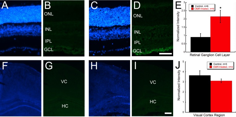
Visual experience during the critical period modulates visual development such that deprivation causes visual impairments while stimulation induces enhancements. This study aimed to determine whether visual stimulation in the form of daily optomotor response (OMR) testing during the mouse critical period (1) improves aspects of visual function, (2) involves retinal mechanisms and (3) is mediated by brain derived neurotrophic factor (BDNF) and dopamine (DA) signaling pathways. We tested spatial frequency thresholds in C57BL/6J mice daily from postnatal days 16 to 23 (P16 to P23) using OMR testing. Daily OMR-treated mice were compared to littermate controls that were placed in the OMR chamber without moving gratings. Contrast sensitivity thresholds, electroretinograms (ERGs), visual evoked potentials, and pattern ERGs were acquired at P21. To determine the role of BDNF signaling, a TrkB receptor antagonist (ANA-12) was systemically injected 2 hours prior to OMR testing in another cohort of mice. BDNF immunohistochemistry was performed on retina and brain sections. Retinal DA levels were measured using high-performance liquid chromatography. Daily OMR testing enhanced spatial frequency thresholds and contrast sensitivity compared to controls. OMR-treated mice also had improved rod-driven ERG oscillatory potential response times, greater BDNF immunoreactivity in the retinal ganglion cell layer, and increased retinal DA content compared to controls. VEPs and pattern ERGs were unchanged. Systemic delivery of ANA-12 attenuated OMR-induced visual enhancements. Daily OMR testing during the critical period leads to general visual function improvements accompanied by increased DA and BDNF in the retina, with this process being requisitely mediated by TrkB activation. These results suggest that novel combination therapies involving visual stimulation and using both behavioral and molecular approaches may benefit degenerative retinal diseases or amblyopia.
Conflict of interest statement
Figures


Similar articles
-
Tau-Driven Neuronal and Neurotrophic Dysfunction in a Mouse Model of Early Tauopathy.J Neurosci. 2016 Feb 17;36(7):2086-100. doi: 10.1523/JNEUROSCI.0774-15.2016. J Neurosci. 2016. PMID: 26888921 Free PMC article.
-
Different effects of valproic acid on photoreceptor loss in Rd1 and Rd10 retinal degeneration mice.Mol Vis. 2014 Nov 4;20:1527-44. eCollection 2014. Mol Vis. 2014. PMID: 25489226 Free PMC article.
-
Retinal and cortical visual acuity in a common inbred albino mouse.PLoS One. 2021 May 28;16(5):e0242394. doi: 10.1371/journal.pone.0242394. eCollection 2021. PLoS One. 2021. PMID: 34048428 Free PMC article.
-
Psychophysical testing in rodent models of glaucomatous optic neuropathy.Exp Eye Res. 2015 Dec;141:154-63. doi: 10.1016/j.exer.2015.06.025. Epub 2015 Jul 2. Exp Eye Res. 2015. PMID: 26144667 Free PMC article. Review.
-
Anatomy and physiology of visual evoked potentials and electroretinograms.Neurol Clin. 1988 Nov;6(4):657-79. Neurol Clin. 1988. PMID: 3070333 Review.
Cited by
-
High-Contrast Stimulation Potentiates the Neurotrophic Properties of Müller Cells and Suppresses Their Pro-Inflammatory Phenotype.Int J Mol Sci. 2022 Aug 3;23(15):8615. doi: 10.3390/ijms23158615. Int J Mol Sci. 2022. PMID: 35955747 Free PMC article.
-
Behavioral Assessment of Visual Function via Optomotor Response and Cognitive Function via Y-Maze in Diabetic Rats.J Vis Exp. 2020 Oct 23;(164):10.3791/61806. doi: 10.3791/61806. J Vis Exp. 2020. PMID: 33165321 Free PMC article.
-
Spontaneous running wheel exercise during pregnancy prevents later neonatal-anoxia-induced somatic and neurodevelopmental alterations.IBRO Neurosci Rep. 2024 Aug 31;17:263-279. doi: 10.1016/j.ibneur.2024.08.008. eCollection 2024 Dec. IBRO Neurosci Rep. 2024. PMID: 39310269 Free PMC article.
-
Effect of amblyopia treatment on macular microvasculature in children with anisometropic amblyopia using optical coherence tomographic angiography.Sci Rep. 2021 Jan 8;11(1):39. doi: 10.1038/s41598-020-79585-4. Sci Rep. 2021. PMID: 33420155 Free PMC article.
-
TrkB Activation during a Critical Period Mimics the Protective Effects of Early Visual Experience on Perception and the Stability of Receptive Fields in Adult Superior Colliculus.J Neurosci. 2019 Jun 5;39(23):4475-4488. doi: 10.1523/JNEUROSCI.2598-18.2019. Epub 2019 Apr 2. J Neurosci. 2019. PMID: 30940716 Free PMC article.
References
-
- Prusky GT, Douglas RM. Developmental plasticity of mouse visual acuity. Eur J Neurosci. 2003;17(1):167–73. . - PubMed
-
- Lunghi C, Burr DC, Morrone MC. Long-term effects of monocular deprivation revealed with binocular rivalry gratings modulated in luminance and in color. Journal of vision. 2013;13(6). Epub 2013/05/03. doi: 10.1167/13.6.1 . - DOI - PubMed
-
- Tschetter WW, Alam NM, Yee CW, Gorz M, Douglas RM, Sagdullaev B, et al. Experience-enabled enhancement of adult visual cortex function. The Journal of neuroscience: the official journal of the Society for Neuroscience. 2013;33(12):5362–6. Epub 2013/03/22. doi: 10.1523/jneurosci.5229-12.2013 ; PubMed Central PMCID: PMCPMC3964778. - DOI - PMC - PubMed
-
- Stafford CA. Critical period plasticity for visual function: definition in monocularly deprived rats using visually evoked potentials. Ophthalmic & physiological optics: the journal of the British College of Ophthalmic Opticians (Optometrists). 1984;4(1):95–100. Epub 1984/01/01. . - PubMed
-
- Sosula L, Glow PH. Increase in number of synapses in the inner plexiform layer of light deprived rat retinae: quantitative electron microscopy. The Journal of comparative neurology. 1971;141(4):427–51. Epub 1971/04/01. doi: 10.1002/cne.901410403 . - DOI - PubMed
Publication types
MeSH terms
Substances
Grants and funding
LinkOut - more resources
Full Text Sources
Other Literature Sources
