. 2018 Feb 6;16(2):e2003885.
doi: 10.1371/journal.pbio.2003885. eCollection 2018 Feb.

Neutrophils kill the parasite Trichomonas vaginalis using trogocytosis

Affiliations

Neutrophils kill the parasite Trichomonas vaginalis using trogocytosis

Frances Mercer et al. PLoS Biol. .

Abstract

T. vaginalis, a human-infective parasite, causes the most common nonviral sexually transmitted infection (STI) worldwide and contributes to adverse inflammatory disorders. The immune response to T. vaginalis is poorly understood. Neutrophils (polymorphonuclear cells [PMNs]) are the major immune cell present at the T. vaginalis-host interface and are thought to clear T. vaginalis. However, the mechanism of PMN clearance of T. vaginalis has not been characterized. We demonstrate that human PMNs rapidly kill T. vaginalis in a dose-dependent, contact-dependent, and neutrophil extracellular trap (NET)-independent manner. In contrast to phagocytosis, we observed that PMN killing of T. vaginalis involves taking "bites" of T. vaginalis prior to parasite death, using trogocytosis to achieve pathogen killing. Both trogocytosis and parasite killing are dependent on the presence of PMN serine proteases and human serum factors. Our analyses provide the first demonstration, to our knowledge, of a mammalian phagocyte using trogocytosis for pathogen clearance and reveal a novel mechanism used by PMNs to kill a large, highly motile target.


Conflict of interest statement

The authors have declared that no competing interests exist.

Figures
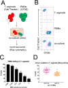
Fig 1
Fig 1. Human PMNs kill T. vaginalis.
(A) Schematic for T. vaginalis cytotoxicity assay: T. vaginalis and PMNs were labelled with CT or CFSE, respectively, and cocultured for 2 hours. Cells were then analyzed by flow cytometry to assess the survival of T. vaginalis. (B) Representative FACS plots of cytotoxicity assay results using an MOI of 0.25 are shown. Surviving T. vaginalis were defined as CT+CFSE. (C) Percent cytotoxicity of T. vaginalis was determined by counting the number of surviving T. vaginalis as outlined in A and B, at various MOIs, defined as parasite:host cell ratio. All data are represented as mean ± SD. (D) Composite results of T. vaginalis cytotoxicity assays, as shown in C, for all 19 donors tested. Each dot represents a different donor. Underlying data can be found in S1 Data and S1, S2, S3, S9 and S10 FCSfiles. CFSE, Carboxyfluorescein succinimidyl ester; CT, Cell Tracker; FACS, fluorescence-activated cell sorting; FCS, fluorescence correlation spectroscopy; MOI, multiplicity of infection; PMN, polymorphonuclear cell.
Fig 2
Fig 2. PMN killing of T. vaginalis is contact-dependent.
(A) and (B) T. vaginalis and PMNs were labelled with CT or CFSE, respectively, and cocultured at the indicated MOI in trans-well plates for 2 hours either together in the bottom well or separate, with T. vaginalis in the top well and PMNs in the bottom well. Cultures were either incubated in the absence or presence of stimulation with 100 nM PMA (lightning bolt symbol) or unlabeled T. vaginalis (white parasites) added at an equivalent MOI in the bottom well. (C) T. vaginalis cytotoxicity assays were performed as described in Fig 1, in the presence of (C) 20,000 U/ml catalase or vehicle control (PBS) or (D) 100 U/ml DNase or vehicle control (HBSS). All data are represented as mean ± SD of triplicate wells and representative of 3 donors and 3 independent experiments. CFSE, Carboxyfluorescein succinimidyl ester; CT, Cell Tracker; HBSS, Hank’s Balanced Salt Solution; MOI, multiplicity of infection; PBS, phosphate-buffered saline; PMA, phorbol-myristate acetate; PMN, polymorphonuclear cell.
Fig 3
Fig 3. PMN killing of T. vaginalis involves engulfment.
T. vaginalis cytotoxicity assay was performed as described in Fig 1, except PMNs were pre-incubated with 2.5 ug/ml Cytochalasin D or vehicle control (DMSO) in panel (A) or 50 ng/ml wortmannin or vehicle control (DMSO) in panel (B) for 20 minutes before T. vaginalis were added. All data are represented as mean ± SD of triplicate wells and representative of 3 donors, and 3 independent experiments. Underlying data can be found in S1 Data. (C) T. vaginalis and PMNs were labelled with CT or CFSE, respectively, and cocultured for 1 hour. Wells were then harvested, fixed with 4% PFA, and analyzed using Imaging Flow Cytometry at a magnification of 63X. Representative images of CFSE+CT+ (Double positives), CFSE-CT+ (T. vaginalis), and CFSE+CT (PMN) populations are shown. BF are bright field images. (D) The total double positives population was analyzed for CT+ spots within CFSE+ cells. (E) Internalization erode score of CT+ signal within CFSE+ cells was determined for the total double positives population. Internalization erode is the ratio of CT+ signal inside of the CFSE+ area versus outside. Data are representative of 3 donors and 3 independent experiments. (F) T. vaginalis were labelled with CT and then incubated at 65 °C for 1 hour and confirmed dead. T. vaginalis were then cocultured with CFSE-labelled PMNs at identical conditions to that shown in Fig 3, and analyzed by imaging flow cytometry. Representative events from the CFSE+CT+ double positive gate are shown. BF are bright field images of double positive cells. BF, bright field; CFSE, Carboxyfluorescein succinimidyl ester; CT, Cell Tracker; MOI, multiplicity of infection; PFA, paraformaldehyde; PMN, polymorphonuclear cell.
Fig 4
Fig 4. PMNs swarm and trogocytose T. vaginalis prior to T. vaginalis death.
(A) Live imaging. Total T. vaginalis surface proteins were covalently linked with Biotin, stained with Streptavidin-Alexa-488 (A488, green), and cocultured with unlabeled PMNs at MOI 0.1 with 10 ug/ml PI in the media. Selected time points of 2D live video at 63X magnification of the interactions are shown. Images are representative of 96 parasite death events performed with PMNs from 11 different human donors. min. = minutes after parasites and PMNs were cocultured. Scale bar = 10 um. (B) and (C) Box and whisker plots showing distribution according to quartiles. (B) 96 videos of T. vaginalis death events from 11 donors’ PMNs were analyzed for the number of PMNs in contact with T. vaginalis before the parasite dies. (C) 96 videos from 11 donors’ PMNs were analyzed for the number of “bites” of T. vaginalis material transferred to PMNs before the parasite dies. (D) Videos in which the first “bite” was observable (19 videos from 7 donors’ PMNs) were analyzed for the time elapsed from when the first bite was taken until PI signal was observed in T. vaginalis. Underlying data can be found in S1 Data. (E) 3D live imaging. T. vaginalis surface was labelled with Alexa-488 as in (A) and cocultured with CT-labelled PMNs (magenta) at MOI 0.1 in the presence of 10 ug/ml PI. Z-stacks spanning 15 um were acquired every 1.5 seconds. Selected time points at 63x of PMN engulfment of T. vaginalis material before parasite death are shown with 3D reconstruction of deconvolved images. Images are clipped midway through the z-axis to visualize the inside of the PMNs. Scale bar = 5 um. White arrow indicates “bite” of T. vaginalis that is uptaken by PMNs. (F) Last timeframe of (E) is shown without any clipping of the z-axis. Data in E and F are representative of at least 3 replicates each from 3 donors and 3 independent experiments. BF, bright field; CT, Cell Tracker; MOI, multiplicity of infection; PI, Propidium Iodide; PMN, polymorphonuclear cell.
Fig 5
Fig 5. Serine proteases are required for trogocytosis and killing of T. vaginalis.
(A) Live imaging was performed identical to that in Fig 4A, except for the pre-incubation of PMNs either with water (vehicle control) or 1-mM AEBSF for 20 minutes prior to addition of parasites. Scale bar = 10 um. (B) Videos of at least 3 parasites bound to PMNs, chosen at random from 3 donors’ PMNs each were analyzed for the number of “bites” of T. vaginalis material transferred to PMNs before parasite death, in the presence of vehicle or 1-mM AEBSF, which were pre-incubated with PMNs for 20 minutes before addition of parasites. The number of bites observed was divided by the number of PMNs present in the swarm, in each case. Total number of parasites analyzed was 15 in the vehicle group and 21 in AEBSF. (C) PMN killing of T. vaginalis at MOI 0.125 (same MOI as panels A and B) was determined using the flow cytometry–based cytolysis assay, in the presence of vehicle or 1-mM AEBSF. Data are represented as mean ± SD of triplicate wells and representative of 3 donors and 3 independent experiments. Underlying data can be found in S1 Data. BF, bright field; MOI, multiplicity of infection; PI, Propidium Iodide; PMN, polymorphonuclear cell.
Fig 6
Fig 6. Human serum factors are required for PMN killing of T. vaginalis.
(A, B) T. vaginalis were stained with 100% human serum, followed by (A) antihuman immunoglobulin light chain kappa or (B) antihuman iC3b, and analyzed with flow cytometry. Green = unstained, red = secondary only (A) or isotype control (B), and blue = fully stained. (C) T. vaginalis cytotoxicity assay was performed as in Fig 1, either in the presence or absence of 10% human serum. (D) T. vaginalis cytotoxicity assay was performed in the presence of 8-ug/ml human IgG Fc fragment or equivalent BSA control. (E) Percent decrease in CT+ events within total CFSE+ events after T. vaginalis cytotoxicity experiments in (C) and (D). All data are represented as mean ± SD of triplicate wells and representative of 3 donors and 3 independent experiments. Underlying data can be found in S1 Data and S5–S8 FCSfiles. BSA, bovine serum albumin; CFSE, Carboxyfluorescein succinimidyl ester; CT, Cell Tracker; Fc, fragment crystallizable; FCS, fluorescence correlation spectroscopy; MOI, multiplicity of infection; PMN, polymorphonuclear cell.

Similar articles

Cited by

References

    1. Schwebke JR, Burgess D (2004) Trichomoniasis. Clin Microbiol Rev 17: 794–803, table of contents. doi: 10.1128/CMR.17.4.794-803.2004 - DOI - PMC - PubMed
    1. Carlton JM, Hirt RP, Silva JC, Delcher AL, Schatz M, et al. (2007) Draft genome sequence of the sexually transmitted pathogen Trichomonas vaginalis. Science 315: 207–212. doi: 10.1126/science.1132894 - DOI - PMC - PubMed
    1. Hirt RP, Sherrard J (2015) Trichomonas vaginalis origins, molecular pathobiology and clinical considerations. Curr Opin Infect Dis 28: 72–79. doi: 10.1097/QCO.0000000000000128 - DOI - PubMed
    1. Kissinger P (2015) Trichomonas vaginalis: a review of epidemiologic, clinical and treatment issues. BMC Infect Dis 15: 307 doi: 10.1186/s12879-015-1055-0 - DOI - PMC - PubMed
    1. Secor WE, Meites E, Starr MC, Workowski KA (2014) Neglected parasitic infections in the United States: trichomoniasis. Am J Trop Med Hyg 90: 800–804. doi: 10.4269/ajtmh.13-0723 - DOI - PMC - PubMed

Substances