

Simulation for competency assessment in vascular and cardiac ultrasound
- PMID: 29409435
- PMCID: PMC6102395
- DOI: 10.1177/1358863X17751656
Simulation for competency assessment in vascular and cardiac ultrasound
Abstract
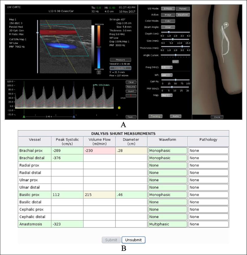
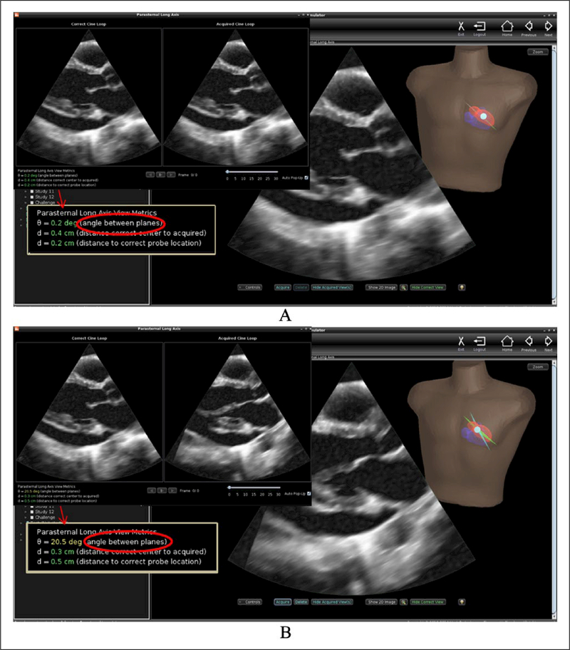
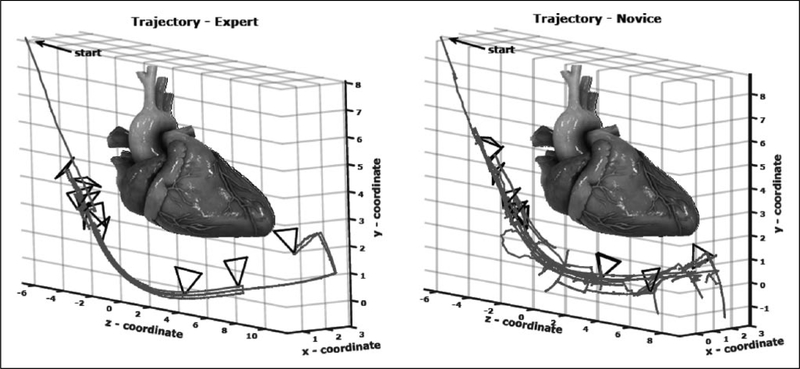
Healthcare providers who use peripheral vascular and cardiac ultrasound require specialized training to develop the technical and interpretive skills necessary to perform accurate diagnostic tests. Assessment of competence is a critical component of training that documents a learner's progress and is a requirement for competency-based medical education (CBME) as well as specialty certification or credentialing. The use of simulation for CBME in diagnostic ultrasound is particularly appealing since it incorporates both the psychomotor and cognitive domains while eliminating dependency on the availability of live patients with a range of pathology. However, successful application of simulation in this setting requires realistic, full-featured simulators and appropriate standardized metrics for competency testing. The principal diagnostic parameter in peripheral vascular ultrasound is measurement of peak systolic velocity (PSV) on Doppler spectral waveforms, and simulation of Doppler flow detection presents unique challenges. The computer-based duplex ultrasound simulator developed at the University of Washington uses computational fluid dynamics modeling and presents real-time color-flow Doppler images and Doppler spectral waveforms along with the corresponding B-mode images. This simulator provides a realistic scanning experience that includes measuring PSV in various arterial segments and applying actual diagnostic criteria. Simulators for echocardiography have been available since the 1990s and are currently more advanced than those for peripheral vascular ultrasound. Echocardiography simulators are now offered for both transesophageal echo and transthoracic echo. These computer-based simulators have 3D graphic displays that provide feedback to the learner and metrics for assessment of technical skill that are based on transducer tracking data. Such metrics provide a motion-based or kinematic analysis of skill in performing cardiac ultrasound. The use of simulation in peripheral vascular and cardiac ultrasound can provide a standardized and readily available method for training and competency assessment.
Keywords: Doppler ultrasound; duplex scanning; echocardiography; medical education; simulation.
Conflict of interest statement
Declaration of conflicting interests
The authors declared the following potential conflicts of interest with respect to the research, authorship, and/or publication of this article: Dr Sheehan is the founder of VentriPoint, Inc., of which she is a major equity holder. VentriPoint markets a product for measuring right heart function, which is not the subject of the present report. Dr Sheehan is also the Founder and President of Sheehan Medical LLC, which markets the transthoracic echocardiography (TTE) simulator that she and co-investigators developed and validated at the University of Washington. Dr Sheehan supports research in medical education by lending TTE simulators from her laboratory at the University of Washington to investigators for up to 6 months. Neither the co-author of the present report nor the University of Washington have involvement in Sheehan Medical LLC, and none receive any benefit from simulator sales.
Figures
References
-
- Institute of Medicine. To Err is Human: Building a Safer Health System. Washington, DC: National Academy Press, 1999.
-
- McGaghie WC, Sajid AW, Miller GE, et al. Competency-Based Curriculum Development in Medical Education. Geneva: World Health Organization, 1978, p.91. - PubMed
-
- Satava RM. Historical review of surgical simulation: A personal perspective. World J Surg 2007; 32: 141–148. - PubMed
-
- Accreditation Council for Graduate Medical Education. Outcome Project: http://njms.rutgers.edu/culweb/medical/documents/ToolboxofAssessmentMeth... (2000, accessed 14 January 2018).
-
- Accreditation Council for Graduate Medical Education and American Board of Medical Specialties Joint Initiative. ACGME Competencies: Suggested Best Methods for Evaluation. Attachment/ Toolbox of Assessment Methods: https://www.partners.org/Assets/Documents/Graduate-Medical-Education/Too... (2000, accessed 14 January 2018).
Publication types
MeSH terms
Grants and funding
LinkOut - more resources
Full Text Sources
Other Literature Sources
