. 2018 Feb 6;18(1):7.
doi: 10.1186/s12896-017-0409-7.

DNA methylome profiling at single-base resolution through bisulfite sequencing of 5mC-immunoprecipitated DNA

Affiliations

DNA methylome profiling at single-base resolution through bisulfite sequencing of 5mC-immunoprecipitated DNA

Zhen Jia et al. BMC Biotechnol. .

Retraction in

Abstract

Background: Detection of DNA methylome at single-base resolution is a significant challenge but promises to shed considerable light on human disease etiology. Current technologies could not detect DNA methylation genome-wide at single-base resolution with small amount of sequencing data and could not avoid detecting the methylation of repetitive elements which are considered as "junk DNA".

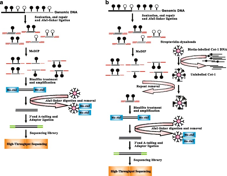
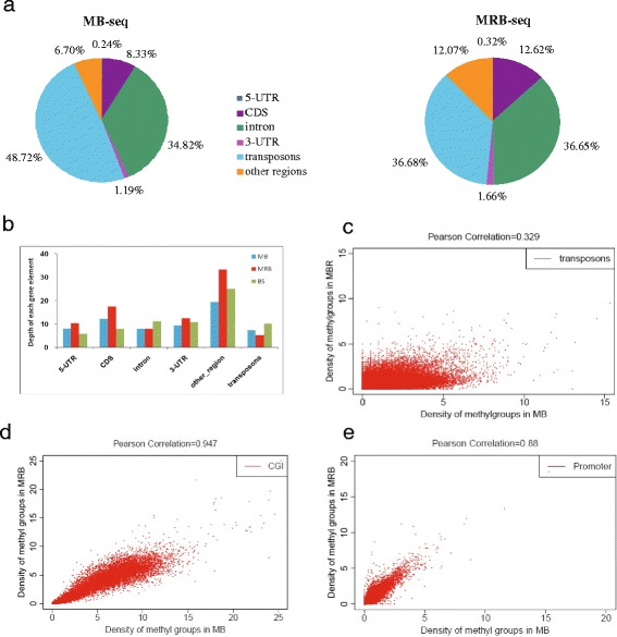
Methods: In this study, we have developed a novel DNA methylome profiling technology named MB-seq with its ability to identify genome-wide 5mC and quantify DNA methylation levels by introduced an assistant adapter AluI-linker This linker can be ligated to sonicated DNA and then be digested after the bisulfite treatment and amplification, which has no effect of MeDIP enrichment. Because many researchers are interested in investigating the methylation of functional regions such as promoters and gene bodies, we have also developed a novel alternative method named MRB-seq, which can be used to investigate the DNA methylation of functional regions by removing the repeats with Cot-1 DNA.

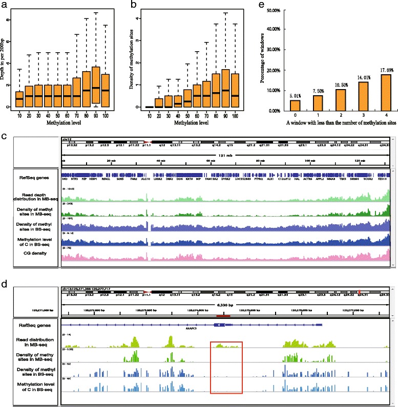
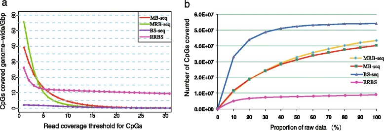
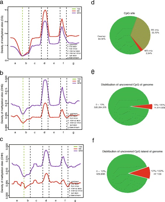
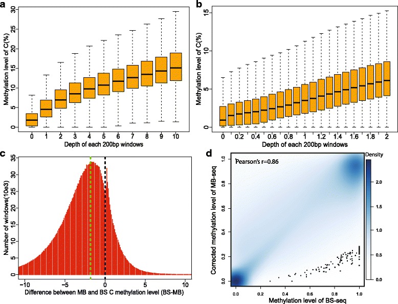
Results: In this study, we have developed MB-seq, a novel DNA methylome profiling technology combining MeDIP-seq with bisulfite conversion, which can precisely detect the 5mC sites and determine their DNA methylation level at single-base resolution in a cost-effective way. In addition, we have developed a new alternative method, MRB-seq (MeDIP-repetitive elements removal-bisulfite sequencing), which interrogates 5mCs in functional regions by depleting nearly half of repeat fragments enriched by MeDIP. Comparing MB-seq and MRB-seq to whole-genome BS-seq using the same batch of DNA from YH peripheral blood mononuclear cells. We found that the sequencing data of MB-seq and MRB-seq almost reaches saturation after generating 7-8 Gbp data, whereas BS-seq requires about 100 Gbp data to achieve the same effect. In comparison to MeDIP-seq and BS-seq, MB-seq offers several key advantages, including single-base resolution, discriminating the methylated sites within a CpG and non-CpG pattern and overcoming the false positive of MeDIP-seq due to the non-specific binding of 5-methylcytidine antibody to genomic fragments.

Conclusion: Our novel developed method MB-seq can accelerate the decoding process of DNA methylation mechanism in human diseases because it requires 7-8 Gbp data to measure human methylome with enough coverage and sequencing depth, affording it a direct and practical application in the study of multiple samples. In addition, we have also provided a novel alternative MRB-seq method, which removes most repetitive sequences and allows researchers to genome-wide characterize DNA methylation of functional regions.

Keywords: DNA methylome; MB-seq; MRB-seq; Novel technology; Single-base resolution.


Conflict of interest statement

Ethics approval and consent to participate

The methods were carried out in accordance with the approved guidelines. This study was approved by written consent from the ethical committee of the Beijing Aviation General Hospital. Relevant informed consent was obtained from all participants.

Consent for publication

Not applicable.

Competing interests

The authors declare that there are no competing interest regarding to publishing this paper.

Figures

Fig. 1
Fig. 1
Schematic outline of the MB-seq and MRB-seq experiment. a Schematic drawing of MB-seq approach. Genomic DNA was randomly fragmented to 100–300 bp and ligated to AluI-linkers with a methylated AluI recognition site close to the T-overhang. The ligated-fragments were captured using methylcytosine antibody, then treated with sodium bisulfite and converted to double stranded DNA by amplification using biotin labelled AluI primers. The AluI-linker was digested and removed by streptavidin coupled beads. The linker-removed sequence was added 3’end A-tailing, then ligated to Illumina multiplexing adaptors following PCR amplification using Illumina paired-end PCR primers. The PCR products of 230–250 bases in length were size-selected on a gel and sequenced on the Illumina platform. b Schematic drawing of MRB-seq approach. Repetitive DNA elements were removed using Cot-1 DNA after MeDIP (based on the MB-seq approach). Cot-1 DNA was labelled with biotin and coupled with streptavidin, and the streptavidin-biotin-Cot-1 DNA was hybridized to enriched methylated DNA fragments via MeDIP to remove repeat fragments. The methylated fragments obtained (single/low copy DNA fragments) were then subjected to sodium bisulfite treatment, PCR amplification, AluI digestion and sequencing library preparation, as per MB-seq
Fig. 2
Fig. 2
False positive exclusion of MeDIP-seq by MB-seq. a The distribution of sequencing depth across different methylation levels in MB-seq. b The distribution of the density of methylation sites across different methylation levels in MB-seq. All information was obtained using a 200 bp window on the genome-wide level. c The distribution of read depth, density of methylation sites in MB-seq and methylation level of 5mC in BS-seq across a randomly genomic region. d Zooming in to a specific region, the red box shows a captured region with no methylated sites, which were nonspecific DNA fragments captured by 5’methylcytosine antibody. e The percentage of windows (200 bp) with less certain methylation sites
Fig. 3
Fig. 3
The genome-wide CpG coverage per Gbp and saturation analysis of BS-seq, MB-seq and MRB-seq. a CpG coverage per Gbp as a function of read coverage threshold for CpGs on the genome-wide level. Coverage of CpGs per Gbp was calculated as (CpGs covered with more than three reads)/all sequencing data in Gbp. b CpG coverage in different amount of sequencing data of BS-seq, MB-seq and MRB-seq
Fig. 4
Fig. 4
Repetitive DNA elements depleting by MRB-seq. a Read disribution of MB-seq and MRB-seq across different genomic features. b Sequencing depth of MB-seq and MRB-seq across different genomic features. MRB-seq successfully depleted repeat sequences and increased the percentage of other genomic features. c-e Scatter plots displaying the correlation of methylation levels between MB-seq and MRB-seq in transposons (c), CGIs (d) and promoters (e). It is shown that, other than in transposons, the repeats-removal process did not significantly affect the methylation level of other genomic features
Fig. 5
Fig. 5
The methylation levels of MB-seq and MRB-seq in gene-associated regions. a Average methylation level of CpG in gene-associated regions using MB-seq and MRB-seq. Gene structure is divided into seven different functional regions and shown on x-axis. The y-axis is the average density of methylation sites in a 200 bp window. The green vertical line shows the mean location of the transcription start sites (TSS). b Average methylation level of CHG in gene-associated regions from MB-seq and MRB-seq. c Average methylation level of CHH in gene-associated regions from MB-seq and MRB-seq. MB-seq and MRB-seq clearly reflect the pattern of methylation level across different functional regions because of the enrichment of methylated non-CpG fragments. d The concordance and difference of CpG sites between MB-seq and BS-seq. e On the genome-wide level, and (f) CpG island-only level, the CpG sites covered solely in the BS-seq dataset were derived from low methylation level
Fig. 6
Fig. 6
Revised CpG methylation level of MB-seq. a Depth within 1–10 X distributed with Methylation level of C on genome. b Distribution of depth within 0–2 X and methylation level of C on genome. c Difference of methylation level C between MB and BS in 200 bp windows (BS-MB). d Correlation between revised methylation level of CG in MB-seq and BS-seq in 200 bp windows on genomewide

Similar articles

Cited by

References

    1. Lister R, Pelizzola M, Dowen RH, Hawkins RD, Hon G, Tonti-Filippini J, Nery JR, Lee L, Ye Z, Ngo QM, et al. Human DNA methylomes at base resolution show widespread epigenomic differences. Nature. 2009;462(7271):315–322. doi: 10.1038/nature08514. - DOI - PMC - PubMed
    1. Cokus SJ, Feng S, Zhang X, Chen Z, Merriman B, Haudenschild CD, Pradhan S, Nelson SF, Pellegrini M, Jacobsen SE. Shotgun bisulphite sequencing of the Arabidopsis genome reveals DNA methylation patterning. Nature. 2008;452(7184):215–219. doi: 10.1038/nature06745. - DOI - PMC - PubMed
    1. Laurent L, Wong E, Li G, Huynh T, Tsirigos A, Ong CT, Low HM, Kin Sung KW, Rigoutsos I, Loring J, et al. Dynamic changes in the human methylome during differentiation. Genome Res. 2010;20(3):320–331. doi: 10.1101/gr.101907.109. - DOI - PMC - PubMed
    1. Robertson KD, Wolffe AP. DNA methylation in health and disease. Nat Rev Genet. 2000;1(1):11–19. doi: 10.1038/35049533. - DOI - PubMed
    1. Suzuki MM, Bird A. DNA methylation landscapes: provocative insights from epigenomics. Nat Rev Genet. 2008;9(6):465–476. doi: 10.1038/nrg2341. - DOI - PubMed

Publication types