Review
. 2018 Feb 6;37(1):20.
doi: 10.1186/s13046-017-0667-1.

Fate of Antibody-Drug Conjugates in Cancer Cells

Affiliations
Review

Fate of Antibody-Drug Conjugates in Cancer Cells

Cécile Chalouni et al. J Exp Clin Cancer Res. .

Abstract

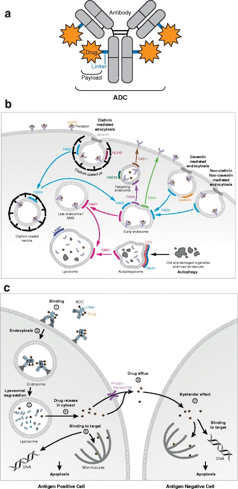
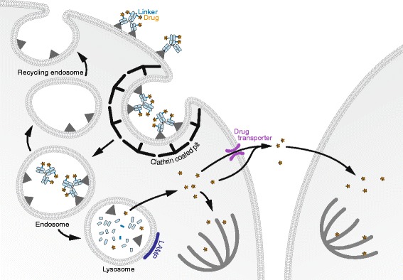
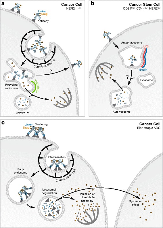
Antibody-Drug Conjugates (ADCs) are a class of cancer therapeutics that combines antigen specificity and potent cytotoxicity in a single molecule as they are comprised of an engineered antibody linked chemically to a cytotoxic drug. Four ADCs have received approval by the Food and Drug Administration (FDA) and the European Medicine Agency (EMA) and can be prescribed for metastatic conditions while around 60 ADCs are currently enrolled in clinical trials. The efficacy of an ADC greatly relies on its intracellular trafficking and processing of its components to trigger tumor cell death. A limited number of studies have addressed these critical processes that both challenge and help foster the design of ADCs. This review highlights those mechanisms and their relevance for future development of ADCs as cancer therapeutics.

Keywords: Antibody-drug conjugates; Endocytic compartments; Endocytosis; Intracellular trafficking.


Conflict of interest statement

Competing interests

The authors declare that they have no competing interests.

Publisher’s Note

Springer Nature remains neutral with regard to jurisdictional claims in published maps and institutional affiliations.

Figures

Fig. 1
Fig. 1
a - ADC structure: An ADC is composed of an antibody coupled to cytotoxic drugs by linkers. b - ADC trafficking and processing classic model: The ADC binds to its surface antigen (1) and the complex is internalized (2), it reaches lysosomes where its linker of the ADC is degraded leading to the release of the drug (3), the drug passes from the intracellular compartment to the cytosol (4), and binds to its target, DNA or tubulin (5) ensue apoptosis. It may also be released into the microenvironment via pumps or passive transfer through the cell membrane (6), capacity to enter a neighbor cancer cell (7) resulting in bystander effect. c- Endocytosis and autophagy pathways
Fig. 2
Fig. 2
Mode of action of brentuximab-vedotin. In Hodgkin lymphoma CD30 positive cells, brentuximab-vedotin is internalized via clathrin coated pits and reaches endosomes. CD30 can recycle or be newly synthesized and re-expressed at the cell surface. The anti-CD30 ADC complex reaches lysosomes where the linker is cleaved by proteases. MMAE molecules traverse the lysosomal membrane and access the cytosol. MMAE can bind the microtubules, triggering depolymerisation leading to cell death. MMAE can traverse the plasma membrane of certain cell lines and exert a toxic effect on neighbor cells (bystander effect)
Fig. 3
Fig. 3
Mode of action of anti-HER2 based ADCs, T-DM1 and the Biparatopic anti-HER2 ADC. a - In HER2 positive cancer cells, T-DM1 is internalized via clathrin coated pits and reaches endosomes where the antibody is degraded leading to the release of DM1. A small fraction of T-DM1 reaches the lysosomes where it can also be degraded. DM1 traverse the lysosomal/endosomal membrane and accesses the cytosol. DM1 can then bind the microtubules, triggering depolymerisation leading to cell death. b - In cancer stem cells, T-DM1 is endocytosed via autophagy. It reaches autophagosomes and later autolysosomes where it is degraded leading to the release of DM1. DM1 accesses the cytosol and inhibits microtubules polymerization leading to cell death. c - The biparatopic anti-HER2 ADC has more efficient trafficking to the lysosomes compared to the monoclonal T-DM1. Its drug tubulysin is liberated in the lysosomes and accesses the cytosol where it can induce the depolymerization of microtubules. In addition, it can exert a bystander effect

Similar articles

Cited by

References

    1. Golay J, Introna M. Mechanism of action of therapeutic monoclonal antibodies: promises and pitfalls of in vitro and in vivo assays. Arch Biochem Biophys. 2012;526(2):146–153. doi: 10.1016/j.abb.2012.02.011. - DOI - PubMed
    1. Sliwkowski MX, Mellman I. Antibody therapeutics in cancer. Science. 2013;341(6151):1192–1198. doi: 10.1126/science.1241145. - DOI - PubMed
    1. Flygare JA, Pillow TH, Aristoff P. Antibody-drug conjugates for the treatment of cancer. Chem Biol Drug Des. 2013;81(1):113–121. doi: 10.1111/cbdd.12085. - DOI - PubMed
    1. Schrama D, Reisfeld RA, Becker JC. Antibody targeted drugs as cancer therapeutics. Nat Rev Drug Discov. 2006;5(2):147–159. doi: 10.1038/nrd1957. - DOI - PubMed
    1. Teicher BA, Chari RV. Antibody conjugate therapeutics: challenges and potential. Clin Cancer Res. 2011;17(20):6389–6397. doi: 10.1158/1078-0432.CCR-11-1417. - DOI - PubMed

MeSH terms