. 2018 Feb 6;9(1):31.
doi: 10.1186/s13287-017-0752-6.

Mesenchymal stromal cell-derived exosome-rich fractionated secretome confers a hepatoprotective effect in liver injury

Affiliations

Mesenchymal stromal cell-derived exosome-rich fractionated secretome confers a hepatoprotective effect in liver injury

Apeksha Damania et al. Stem Cell Res Ther. .

Abstract

Background: Mesenchymal stromal cells (MSCs) are an attractive therapeutic agent in regenerative medicine. Recently, there has been a paradigm shift from differentiation of MSCs to their paracrine effects at the injury site. Several reports elucidate the role of trophic factors secreted by MSCs toward the repair of injured tissues. We hypothesize that fractionating the MSC secretome will enrich exosomes containing soluble bioactive molecules, improving its therapeutic potential for liver failure.

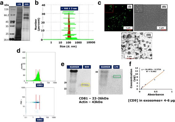
Methods: Rat bone marrow MSCs were isolated and the conditioned media filtered, concentrated and ultracentrifuged to generate fractionated secretome. This secretome was characterized for the presence of exosomes and recovery from liver injury assessed in in-vitro liver injury models. The results were further validated in vivo.

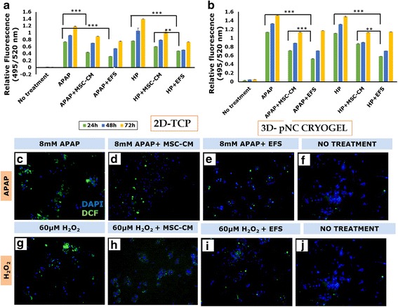
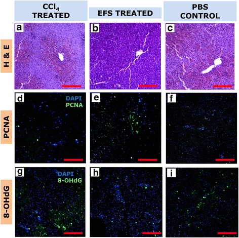
Results: Studies on in-vitro liver injury models using acetaminophen and hydrogen peroxide show better cell recovery and reduced cytotoxicity in the presence of fractionated as opposed to unfractionated secretome. Further, the cells showed reduced oxidative stress in the presence of fractionated secretome, suggesting a potential antioxidative effect. These results were further validated in vivo in liver failure models, wherein improved liver regeneration in the presence of fractionated secretome (0.819 ± 0.035) was observed as compared to unfractionated secretome (0.718 ± 0.042).

Conclusions: The work presented is a proof of concept that fractionating the secretome enriches certain bioactive molecules involved in the repair and recovery of injured liver tissue. Exosome enriched mesenchymal stromal cell-derived fractionated secretome potentiates recovery upon injection in injured liver.

Keywords: Cryogel; Exosomes; Liver; Secretome; Stromal cells.


Conflict of interest statement

Ethics approval and consent to participate

Isolation of bone marrow-derived MSCs and development of ALF models were carried out using protocols approved by the Institute Animal Ethics Committee (IITK/IAEC/2014/1023 and IITK/IAEC/2014/1022, respectively) of IIT Kanpur, under the Committee for the Purpose of Control and Supervision of Experiments on Animals (CPCSEA), Government of India. All methods were performed in accordance with relevant guidelines and regulations of this committee.

Consent for publication

Not applicable.

Competing interests

The authors declare that they have no competing interests.

Publisher’s Note

Springer Nature remains neutral with regard to jurisdictional claims in published maps and institutional affiliations.

Figures

Fig. 1
Fig. 1
Schematic representation for generation of fractionated secretome. MSC mesenchymal stromal cell, CM conditioned medium, PBS phosphate buffered saline
Fig. 2
Fig. 2
Characterization of MSC fractionated secretome. SDS-PAGE gel for MSC cell lysate (LYS) and exosome-rich fractionated MSC secretome (EXO) (a). DLS data showing average z-value representative of average particle size (b). Confocal microscopic image at lower (×20) and higher (×100) magnification (inset) (scale bar: 1 μm) (c): (i) scanning electron microscopic image (scale bar: 1 μm), (ii) transmission electron microscopic image (scale bar: 1 μm) and (iii) for EFS. Flow cytometric analysis for presence of exosomal marker CD63 in the fractionated secretome (d). Western blot analysis showing presence of exosomal marker CD81 (e). Standard curve for sandwich ELISA performed to confirm presence of exosomal marker CD9 in the fractionated MSC secretome (f). FSC forward scatter
Fig. 3
Fig. 3
Effect of EFS on viability of liver cells. Effect of different concentrations of exosome-rich fractionated secretome (EFS) on viability of HepG2 cells over a period of 72 h (a). MTT assay to check effect of fractionated secretome and unfractionated secretome on viability of injury-induced liver cells in 2D tissue culture plate (2D-TCP) (b) and 3D cryogel scaffold (3D-pNC CRYOGEL) (c). LDH activity of acetaminophen (APAP) and hydrogen peroxide (HP) injury-induced liver cells before and after treatment with unfractionated MSC secretome (MSC-CM) and fractionated MSC secretome (EFS) (d). Statistical analysis: n = 3, **p < 0.01. 2D two dimensional, 3D three dimensional, MSC mesenchymal stromal cell, CM conditioned medium, LDH lactate dehydrogenase
Fig. 4
Fig. 4
Fluorescent microscopic analysis of the effect of EFS on liver cell viability in injury conditions. Live–dead staining using FDA and PI 24 and 48 h post treatment with 8 mM acetaminophen (APAP) (a and e, respectively), 8 mM APAP in presence of unfractionated MSC secretome (b and f, respectively), 8 mM APAP in presence of fractionated MSC secretome (c and g, respectively) and without any treatment (d and h, respectively). Live–dead staining using FDA and PI 24 and 48 h post treatment with 60 μM hydrogen peroxide (H2O2) (i and m, respectively), 60 μM H2O2 in presence of unfractionated MSC secretome (j and n, respectively), 60 μM H2O2 in presence of fractionated MSC secretome (k and o, respectively) and without any treatment (l and p, respectively). Scale bar for all microscopic images: 100 μm. MSC mesenchymal stromal cell, CM conditioned medium, EFS exosome-rich fractionated secretome
Fig. 5
Fig. 5
Effect of EFS on oxidative stress induced during liver injury. Quantitative DCFDA assay for reactive oxygen species (ROS) released by liver cells post treatment with acetaminophen (APAP) and hydrogen peroxide (HP) in the presence of unfractionated MSC secretome (MSC-CM) and fractionated MSC secretome (EFS) in 2D tissue culture plate (2D-TCP) (a) and 3D cryogel scaffold (3D-pNC CRYOGEL) (b). Qualitative DCFDA assay for ROS 24 h post treatment with 8 mM APAP (c), 8 mM APAP in presence of unfractionated MSC secretome (d), 8 mM APAP in presence of fractionated MSC secretome (e) and without any treatment (f). Qualitative DCFDA assay for ROS 24 h post treatment with 60 μM hydrogen peroxide (H2O2) (g), 60 μM H2O2 in presence of unfractionated MSC secretome (h), 60 μM H2O2 in presence of fractionated MSC secretome (i) and without any treatment (j). Scale bar for all microscopic images: 100 μm. Statistical analysis: n = 3, **p < 0.01, ***p < 0.001. MSC mesenchymal stromal cell, CM conditioned medium, EFS exosome-rich fractionated secretome, 2D two dimensional, 3D three dimensional
Fig. 6
Fig. 6
Effect of EFS on liver regeneration and recovery in ischemic/reperfusion model of liver failure. Liver regeneration rate of untreated rodent model (defect), model treated with unfractionated MSC secretome (defect + MSC-CM) and model treated with fractionated MSC secretome (defect + EFS). Statistical analysis: n = 3 (control); n = 5 (models), *p < 0.05, **p < 0.01 (a). AST (b), ALT (c), bilirubin (d) and albumin (e) levels in untreated liver failure models (PHx), models treated with unfractionated secretome (PHx + MSC-CM) and models treated with fractionated secretome (PHx + EFS). Statistical analysis: n = 3, *p < 0.05, **p < 0.01, #ns. MSC mesenchymal stromal cell, CM conditioned medium, EFS exosome-rich fractionated secretome, AST aspartate transaminase, ALT alanine transaminase, PHx partial hepatectomy
Fig. 7
Fig. 7
Effect of EFS on recovery in carbon tetrachloride-induced liver injury model. ALT, AST, bilirubin and albumin levels in liver failure model before treatment (CCl4 treated) and CCl4 models treated with EFS (EFS treated) and treated with PBS (PBS control) at regular time intervals post injection. Statistical analysis: n = 3, *p < 0.05, **p < 0.01, ***p < 0.001, #ns. AST aspartate transaminase, ALT alanine transaminase, CCl4 carbon tetrachloride, EFS exosome-rich fractionated secretome, PBS phosphate buffered saline
Fig. 8
Fig. 8
Effect of EFS on hepatocyte proliferation and oxidative stress in carbon tetrachloride-induced liver injury. Hematoxylin and eosin (H&E) staining (a–c), PCNA expression (d–f) and 8-OHdG expression (g–i) of untreated (CCl4 treated), EFS (EFS treated) and PBS treated (PBS control) injury models 72 h post injection. Scale bar for all microscopic images: 100 μm. CCl4 Carbon tetrachloride; EFS Exosome-rich fractionated secretome; PBS Phosphate buffered saline; PCNA Proliferating cell nuclear antigen; 8-OHdG 8-hydroxy-2' -deoxyguanosine; DAPI 4',6-diamidino-2-phenylindole

References

    1. Michalopoulos GK, DeFrances MC. Liver regeneration. Science. 1997;276(5309):60–6. doi: 10.1126/science.276.5309.60. - DOI - PubMed
    1. Taub R. Liver regeneration: from myth to mechanism. Nat Rev Mol Cell Biol. 2004;5(10):836–47. doi: 10.1038/nrm1489. - DOI - PubMed
    1. Fausto N. Liver regeneration and repair: hepatocytes, progenitor cells, and stem cells. Hepatology. 2004;39(6):1477–87. doi: 10.1002/hep.20214. - DOI - PubMed
    1. Michalopoulos GK. Liver regeneration. J Cell Physiol. 2007;213(2):286–300. doi: 10.1002/jcp.21172. - DOI - PMC - PubMed
    1. Canbay A, Tacke F, Hadem J, Trautwein C, Gerken G, Manns MP. Acute Liver Failure: A Life-Threatening Disease. Dtsch Arztebl Int. 2011;108(42):714–20. http://doi.org/10.3238/arztebl.2011.0714. - DOI - PMC - PubMed

Publication types

MeSH terms