. 2018 May 18;18(2):135-147.
doi: 10.3727/105221618X15174108894682. Epub 2018 Feb 6.

The Effect of Selective c-MET Inhibitor on Hepatocellular Carcinoma in the MET-Active, β-Catenin-Mutated Mouse Model

Affiliations

The Effect of Selective c-MET Inhibitor on Hepatocellular Carcinoma in the MET-Active, β-Catenin-Mutated Mouse Model

Na Zhan et al. Gene Expr. .

Abstract

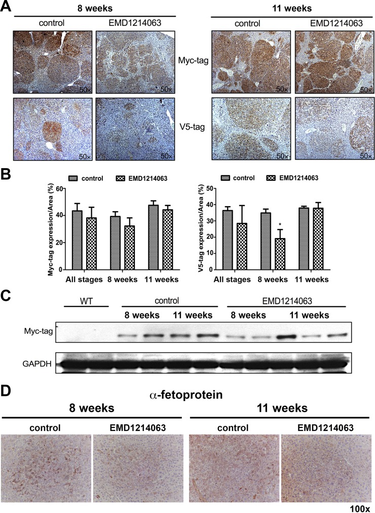
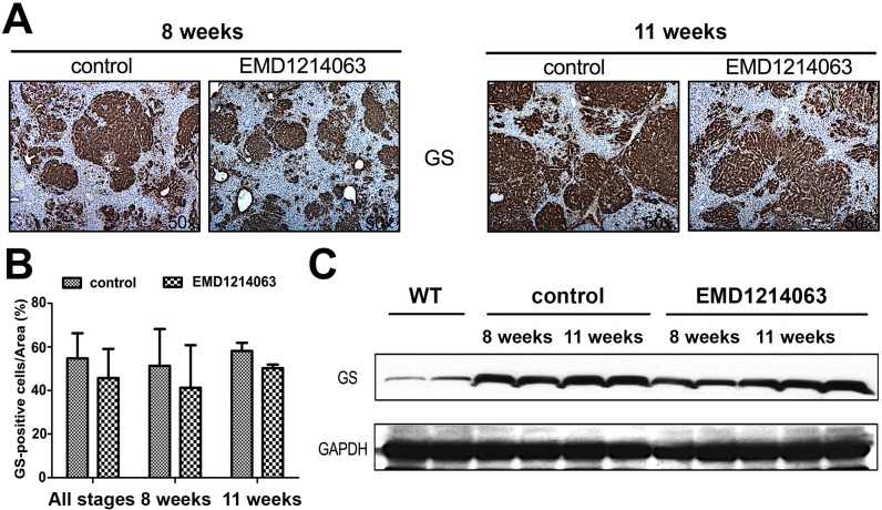
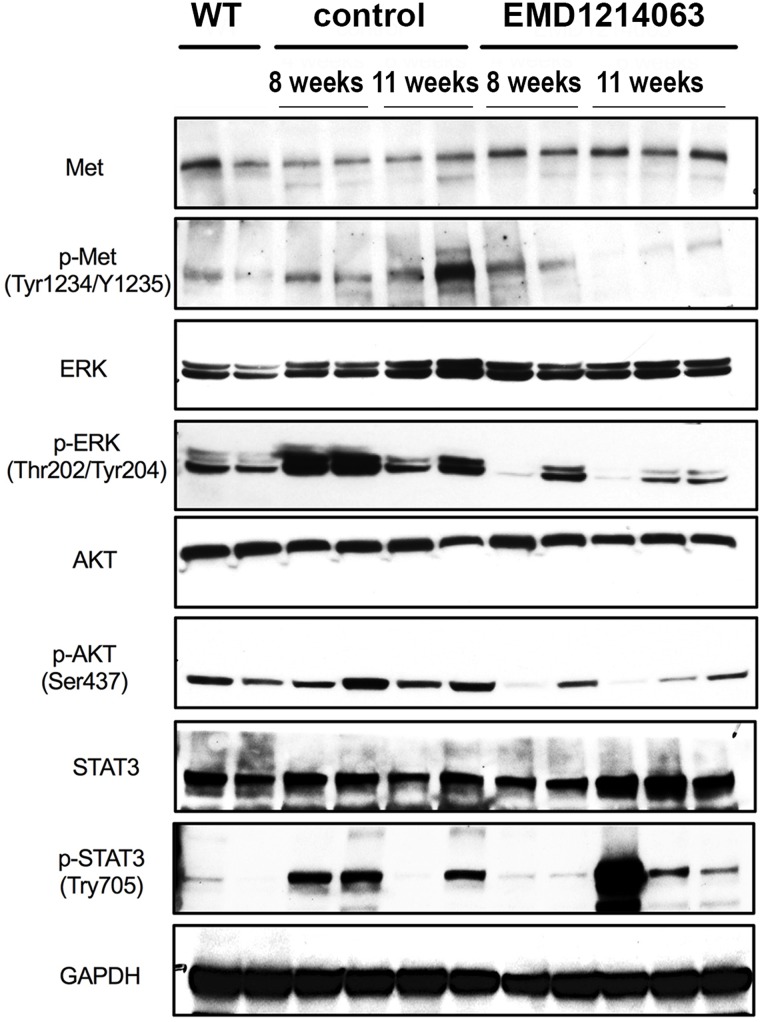
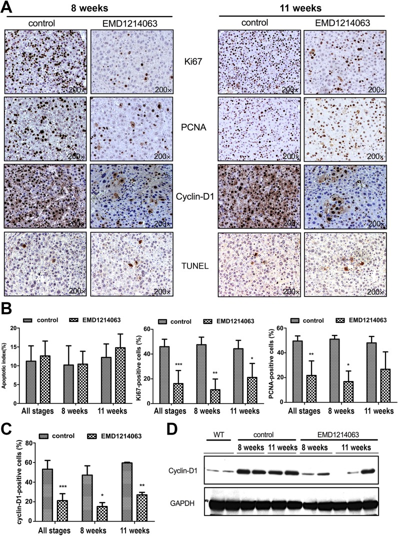
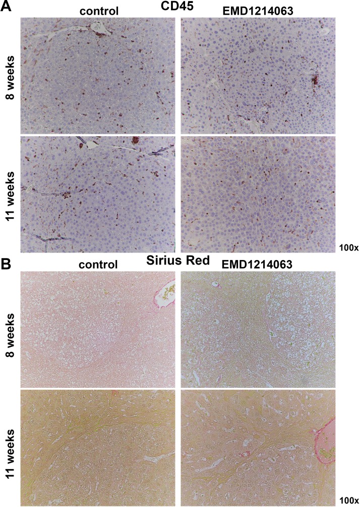
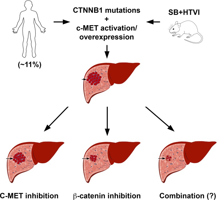
Simultaneous mutations in CTNNB1 and activation of c-MET occur in 9%-12.5% of patients with hepatocellular carcinoma (HCC). Coexpression of c-MET-V5 and mutant β-catenin-Myc in mouse liver by sleeping beauty transposon/transposase and hydrodynamic tail vein injection (SB-HTVI) led to the development of HCC with 70% molecular identity to the clinical subset. Using this model, we investigated the effect of EMD1214063, a highly selective c-MET inhibitor. Five weeks after SB-HTVI when tumors were established, EMD1214063 (10 mg/kg) was administered by gastric gavage as a single agent on 5-day-on/3-day-off schedule, compared to vehicle only control. Mice were harvested at 8 or 11 weeks posttreatment. Decreased p-MET, p-AKT, p-STAT3, and p-ERK proved in vivo efficacy of EMD1214063. We observed lower Ki-67, PCNA, V5-tag, and cyclin D1 after EMD1214063 treatment only at 8 weeks. Overall, no significant differences were observed in tumor burden between the groups, although EMD1214063 marginally but significantly improved overall survival by 1.5-2 weeks. Tumors remained α-fetoprotein+, did not show any differences in inflammation, and lacked fibrosis in either group. In conclusion, c-MET inhibition alone had a minor effect on Met-β-catenin HCC at the early stages of HCC development. Thus, a single therapy with the c-MET inhibitor will be insufficient for sustained response in Met-β-catenin HCC requiring assessment of additional combinations.


Conflict of interest statement

The authors declare no conflicts of interests.

Figures

Figure 1
Figure 1
A marginal improvement in overall survival but lack of any significant effect of treatment with the c-MET inhibitor EMD1214063 for 8 or 11 weeks on tumor growth in the Met-β-catenin model. (A) Schematic illustrating the overall experimental design of the study. (B) Gross liver images show comparable morphology between the control and EMD1241063 groups, although this group showed a somewhat healthy appearance at 8 weeks as shown by lack of relative nodularity. (C) No significant difference in liver weight/body weight (LW/BW) ratio as an indicator of overall tumor burden between the control and EMD1214063 treatment groups overall or at 8 or 11 weeks individually [average ± standard deviation (SD)]. (D) Kaplan–Meier survival curve showing EMD1241063 treatment causing marginal but significant improvement in the overall survival by 1.5–2 weeks compared to the control treatment in the Met-β-catenin model (p = 0.0396). This was based on time to morbidity as indicated by greater than 3 g of liver weight due to excessive tumor burden. (E) Hematoxylin and eosin (H&E) staining of representative liver sections from the EMD1214063 group versus controls shows comparable microscopic foci.
Figure 2
Figure 2
EMD1214063 treatment of the Met-β-catenin model temporally decreases V5-tag (c-MET) but not Myc-tag (β-catenin). (A) Microscopic foci show positivity for both Myc-tag and V5-tag in the control group at both 8 and 11 weeks. EMD1214063 treatment did not have any effect on staining for Myc-tag. Staining for V5-tag showed smaller and fainter positive tumor foci after 8 weeks of EMD1214063, but no difference was observed at 11 weeks. (B) Quantification of immunohistochemistry (IHC) verified lack of significant differences in Myc-tag but showed a significant decrease in V5-tag staining (*p = 0.0465) after EMD1214063 treatment for 8 weeks only. (C) Western blot for Myc-tag showed decrease in Myc-tag levels at 8 weeks of EMD1214063 treatment only. GAPDH shows comparable loading in all lanes. (D) IHC for α-fetoprotein shows tumors to be positive in both control- and EMD1214063-treated group at both 8 and 11 weeks.
Figure 3
Figure 3
EMD1214063 does not impact β-catenin signaling in the Met-β-catenin model. (A) Microscopic foci show positivity for glutamine synthetase (GS) in the control- and EMD1214063-treated groups at both 8 and 11 weeks. (B) Quantification of immunostaining showed lack of any differences in the control and experimental group at either or both time points after EMD1214063 treatment. (C) EMD1214063 treatment showed a marginal decrease in total GS levels by Western blot analysis at only 8 weeks. GAPDH shows comparable loading in all lanes.
Figure 4
Figure 4
EMD1214063 inhibits c-MET signaling in the Met-β-catenin hepatocellular carcinoma (HCC) mouse model. Western blot analysis using whole-liver lysates from control- and EMD1214063-treated groups at 8 and 11 weeks shows a notable decrease in the levels of p-Met (Tyr1234/1235), p-ERK1/2 (Thr202/Tyr204), p-AKT (Ser473), and p-STAT3 (Tyr705) levels in the EMD1214063-treated group. GAPDH shows comparable loading in all lanes.
Figure 5
Figure 5
EMD1214063 reduces cell proliferation after 8 and 11 weeks of treatment of Met-β-catenin mice, which is associated with a decrease in cyclin D1 levels. (A) IHC for Ki-67 and PCNA shows decrease in the EMD1214063 treatment group compared with the control group at both 8 and 11 weeks after treatment. Cyclin D1 levels were also reduced. No change in the number of TUNEL+ cells was evident after EMD1214063 treatment at either time point. (B) Quantification of TUNEL, Ki-67, and PCNA immunohistochemistry shows insignificant difference in apoptosis, but significant differences in Ki-67 at combined time points (***p = 0.0004), at 8 weeks (**p = 0.0096), and at 11 weeks (*p = 0.042), as well as PCNA at combined time points (**p = 0.0016) or at 8 weeks (*p = 0.0225). (C) Quantification of cyclin D1 staining showed a significant difference in EMD1214063-treated samples compared to controls at both times combined (***p < 0.0001), at 8 weeks (*p = 0.0330), and at 11 weeks (**p = 0.0028). (D) Decrease in levels of cyclin D1 after EMD1214063 was verified by Western blots as well. GAPDH shows comparable loading in all lanes.
Figure 6
Figure 6
EMD1214063 does not impact inflammation or fibrosis at 8 weeks or 11 weeks of treatment of Met-β-catenin mice. (A) IHC for CD45 shows comparable inflammatory cells both within and outside the tumor foci at 8 weeks or 11 weeks in controls or EMD1214063 treatment group. (B) Sirius Red staining shows lack of any hepatic fibrosis in control-treated Met-β-catenin mice at either 8 or 11 weeks. No differences between controls and EMD1214063 treatment were evident at either time points.
Figure 7
Figure 7
Cartoon depicting relative response of HCC in the Met-β-catenin mice to c-MET or β-catenin suppression. Eleven percent of all human HCCs shows concomitant β-catenin mutations and c-MET overexpression or activation. Coexpression of these two proto-oncogenes in mouse liver leads to HCC, which molecularly resembles the subset of human HCC with simultaneous β-catenin mutations and c-MET activation. When treated with c-MET inhibitors after tumors were established, only a marginal effect on overall HCC burden was observed. However, suppression of β-catenin led to a profound response, and tumors were notably eliminated in a predominant subset of mice. It is anticipated that HCC in this model may respond even more profoundly to combined c-MET and β-catenin suppression, although it has not been directly investigated yet.

Similar articles

Cited by

References

    1. El-Serag HB. Hepatocellular carcinoma. N Engl J Med. 2011;365(12):1118–27. - PubMed
    1. Jemal A, Bray F, Center MM, Ferlay J, Ward E, Forman D. Global cancer statistics. CA Cancer J Clin. 2011;61(2):69–90. - PubMed
    1. 2017 Cancer stat facts: Liver and intrahepatic bile duct cancer. Available at https://seer.cancer.gov/statfacts/html/livibd.html
    1. Golabi P, Fazel S, Otgonsuren M, Sayiner M, Locklear CT, Younossi ZM. Mortality assessment of patients with hepatocellular carcinoma according to underlying disease and treatment modalities. Medicine (Baltimore) 2017;96(9):e5904. - PMC - PubMed
    1. Bruix J, Qin S, Merle P, Granito A, Huang YH, Bodoky G, Pracht M, Yokosuka O, Rosmorduc O, Breder V, Gerolami R, Masi G, Ross PJ, Song T, Bronowicki JP, Ollivier-Hourmand I, Kudo M, Cheng AL, Llovet JM, Finn RS, LeBerre MA, Baumhauer A, Meinhardt G, Han G; RESORCE Investigators. Regorafenib for patients with hepatocellular carcinoma who progressed on sorafenib treatment (RESORCE): A randomised, double-blind, placebo-controlled, phase 3 trial. Lancet 2017;389(10064):56–66. - PubMed

Publication types

MeSH terms