

Structure and collagen crimp patterns of functionally distinct equine tendons, revealed by quantitative polarised light microscopy (qPLM)
- PMID: 29409868
- PMCID: PMC5894809
- DOI: 10.1016/j.actbio.2018.01.034
Structure and collagen crimp patterns of functionally distinct equine tendons, revealed by quantitative polarised light microscopy (qPLM)
Abstract
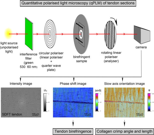
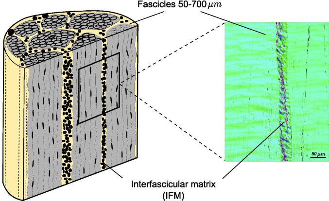
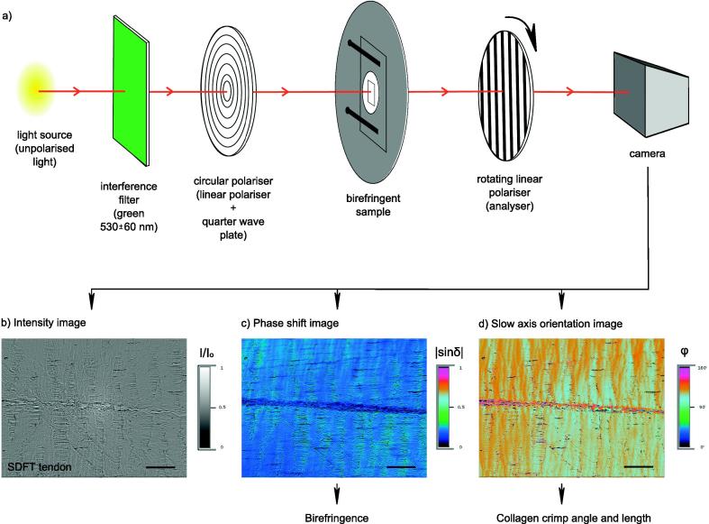
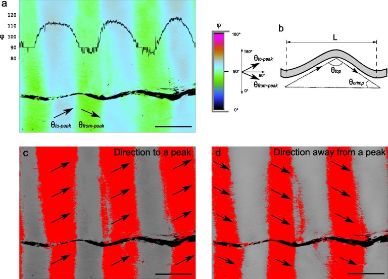
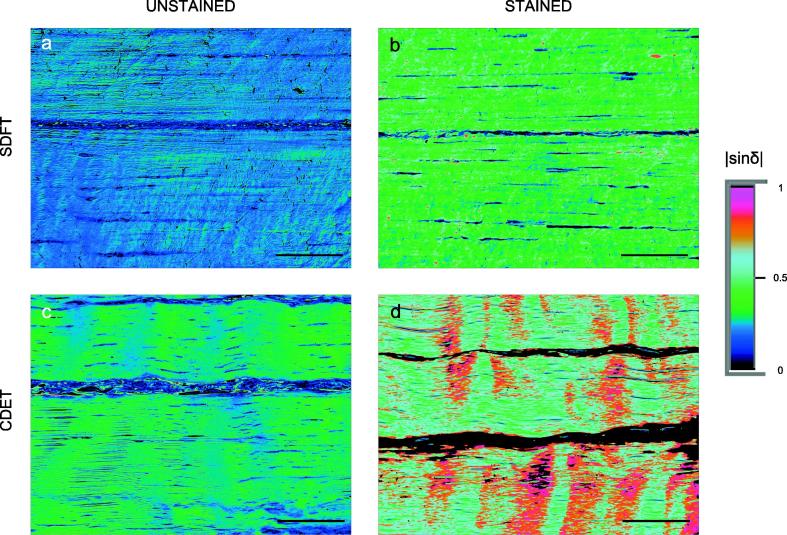
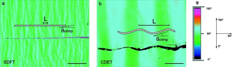
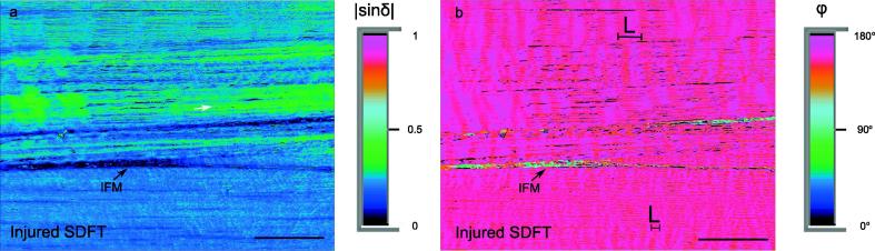
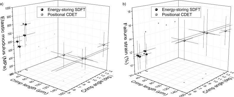
Structure-function relationships in tendons are directly influenced by the arrangement of collagen fibres. However, the details of such arrangements in functionally distinct tendons remain obscure. This study demonstrates the use of quantitative polarised light microscopy (qPLM) to identify structural differences in two major tendon compartments at the mesoscale: fascicles and interfascicular matrix (IFM). It contrasts functionally distinct positional and energy storing tendons, and considers changes with age. Of particular note, the technique facilitates the analysis of crimp parameters, in which cutting direction artefact can be accounted for and eliminated, enabling the first detailed analysis of crimp parameters across functionally distinct tendons. IFM shows lower birefringence (0.0013 ± 0.0001 [-]), as compared to fascicles (0.0044 ± 0.0005 [-]), indicating that the volume fraction of fibres must be substantially lower in the IFM. Interestingly, no evidence of distinct fibre directional dispersions between equine energy storing superficial digital flexor tendons (SDFTs) and positional common digital extensor tendons (CDETs) were noted, suggesting either more subtle structural differences between tendon types or changes focused in the non-collagenous components. By contrast, collagen crimp characteristics are strongly tendon type specific, indicating crimp specialisation is crucial in the respective mechanical function. SDFTs showed much finer crimp (21.1 ± 5.5 µm) than positional CDETs (135.4 ± 20.1 µm). Further, tendon crimp was finer in injured tendon, as compared to its healthy equivalents. Crimp angle differed strongly between tendon types as well, with average of 6.5 ± 1.4° in SDFTs and 13.1 ± 2.0° in CDETs, highlighting a substantially tighter crimp in the SDFT, likely contributing to its effective recoil capacity.
Statement of significance: This is the first study to quantify birefringence in fascicles and interfascicular matrix of functionally distinct energy storing and positional tendons. It adopts a novel method - quantitative polarised light microscopy (qPLM) to measure collagen crimp angle, avoiding artefacts related to the direction of histological sectioning, and provides the first direct comparison of crimp characteristics of functionally distinct tendons of various ages. A comparison of matched picrosirius red stained and unstained tendons sections identified non-homogenous staining effects, and leads us to recommend that only unstained sections are analysed in the quantitative manner. qPLM is successfully used to assess birefringence in soft tissue sections, offering a promising tool for investigating the structural arrangements of fibres in (soft) tissues and other composite materials.
Keywords: Birefringence; Collagen crimp; Fascicles; Interfascicular matrix (endotenon); Quantitative polarised light microscopy (qPLM); Tendon.
Copyright © 2018 Acta Materialia Inc. Published by Elsevier Ltd. All rights reserved.
Figures
References
-
- Handsfield G.G., Slane L.C., Screen H.R.C. Nomenclature of the tendon hierarchy: an overview of inconsistent terminology and a proposed size-based naming scheme with terminology for multi-muscle tendons. J. Biomech. 2016;49:3122–3124. - PubMed
-
- Karunaratne A., Boyde A., Esapa C.T., Hiller J., Terrill N.J., Brown S.D.M., Cox R.D., Thakker R.V., Gupta H.S. Symmetrically reduced stiffness and increased extensibility in compression and tension at the mineralized fibrillar level in rachitic bone. Bone. 2013;52:689–698. - PubMed
Publication types
MeSH terms
Substances
Grants and funding
LinkOut - more resources
Full Text Sources
Other Literature Sources
Medical
Molecular Biology Databases
Research Materials
