

Presumed structural and functional neural recovery after long-term abstinence from cocaine in male military veterans
- PMID: 29410011
- PMCID: PMC5880688
- DOI: 10.1016/j.pnpbp.2018.01.024
Presumed structural and functional neural recovery after long-term abstinence from cocaine in male military veterans
Abstract
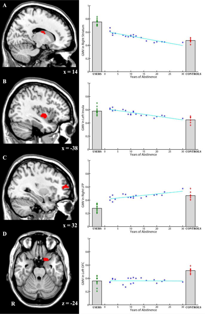
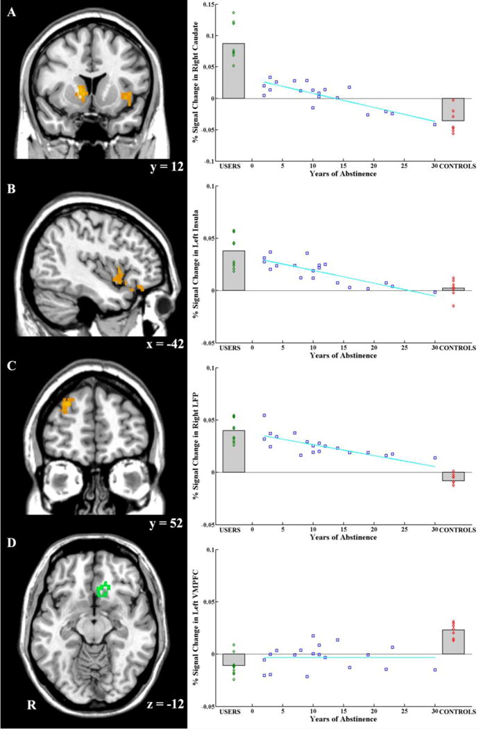
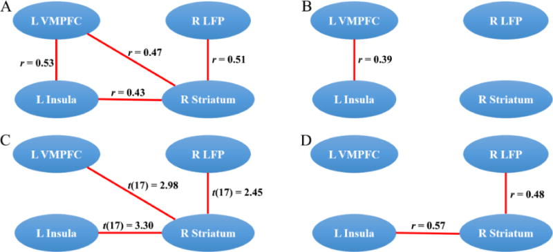
Cumulative evidence suggests that cocaine use could alter the structure and function of different brain systems. However, the extent to which the altered brain structure and function possibly recover over time after cocaine abstinence remains less clear. The present study examines 39 male military veterans with different stages of cocaine addiction and long-term abstinence (from 1 year up to 30 years) and evaluates plausible changes in brain structure and function of specific brain regions that sub-serve addictions. These include the striatum that is involved in cocaine reward; the lateral prefrontal cortex (especially the dorsolateral PFC) that plays a major role in inhibitory control; the insula, which has been implicated in craving; and the medial orbitofrontal (OFC) and ventromedial prefrontal cortex (VMPFC) shown to play key roles in foresight and decision-making. The results suggest that there are differences in both brain structure (gray matter volume, GMV) and function between cocaine USERS and CONTROLS, with USERS showing plausible relative strengthening in neural systems for processing reward and craving, and relative weakening in neural systems involved in inhibitory control and decision-making. Examination of possible neural changes after abstinence suggests that presumed recovery occurs mostly in neural systems related to reward, craving, and inhibitory control, but to a lesser extent in neural systems related to decision-making. Given the limitations of the data in terms of a small sample size, as well as the lack of certainty about occasional use in the abstinent group, these results may be considered as preliminary. However, they are compelling in that they suggest that male military veterans cocaine USERS are indefinitely at a higher risk compared to CONTROLS for making lapses in judgment and decision-making leading to possible relapse, if reward salience and craving become more intense. Understanding the neurobiology of long-term cocaine abstinence in vulnerable populations and beyond could help devising better therapeutic strategies that prevent relapse.
Keywords: Abstinence; Cocaine addiction; Functional connectivity; VBM; fMRI.
Copyright © 2018 Elsevier Inc. All rights reserved.
Conflict of interest statement
The authors declare no conflict of interest.
Figures
Similar articles
-
Bouncing back: Brain rehabilitation amid opioid and stimulant epidemics.Neuroimage Clin. 2019;24:102068. doi: 10.1016/j.nicl.2019.102068. Epub 2019 Nov 5. Neuroimage Clin. 2019. PMID: 31795056 Free PMC article. Review.
-
Cocaine addiction is associated with abnormal prefrontal function, increased striatal connectivity and sensitivity to monetary incentives, and decreased connectivity outside the human reward circuit.Addict Biol. 2017 May;22(3):844-856. doi: 10.1111/adb.12356. Epub 2016 Jan 19. Addict Biol. 2017. PMID: 26786150
-
Salience and default mode network dysregulation in chronic cocaine users predict treatment outcome.Brain. 2017 May 1;140(5):1513-1524. doi: 10.1093/brain/awx036. Brain. 2017. PMID: 28334915 Free PMC article.
-
Brain Volume Correlates with Duration of Abstinence from Substance Abuse in a Region-Specific and Substance-Specific Manner.Biol Psychiatry Cogn Neurosci Neuroimaging. 2017 Oct;2(7):626-635. doi: 10.1016/j.bpsc.2017.03.011. Biol Psychiatry Cogn Neurosci Neuroimaging. 2017. PMID: 29308437 Free PMC article.
-
The alcoholic brain: neural bases of impaired reward-based decision-making in alcohol use disorders.Neurol Sci. 2018 Mar;39(3):423-435. doi: 10.1007/s10072-017-3205-1. Epub 2017 Nov 29. Neurol Sci. 2018. PMID: 29188399 Review.
Cited by
-
Bouncing back: Brain rehabilitation amid opioid and stimulant epidemics.Neuroimage Clin. 2019;24:102068. doi: 10.1016/j.nicl.2019.102068. Epub 2019 Nov 5. Neuroimage Clin. 2019. PMID: 31795056 Free PMC article. Review.
-
A brainnetome atlas-based methamphetamine dependence identification using neighborhood component analysis and machine learning on functional MRI data.Front Cell Neurosci. 2022 Sep 27;16:958437. doi: 10.3389/fncel.2022.958437. eCollection 2022. Front Cell Neurosci. 2022. PMID: 36238830 Free PMC article.
-
Food-Specific Inhibitory Control Mediates the Effect of Disgust Sensitivity on Body Mass Index.Front Psychol. 2019 Oct 22;10:2391. doi: 10.3389/fpsyg.2019.02391. eCollection 2019. Front Psychol. 2019. PMID: 31695662 Free PMC article.
-
Reduced thalamic resting-state functional connectivity and impaired cognition in acute abstinent heroin users.Hum Brain Mapp. 2021 May;42(7):2077-2088. doi: 10.1002/hbm.25346. Epub 2021 Jan 18. Hum Brain Mapp. 2021. PMID: 33459459 Free PMC article.
-
Partial recovery of the left DLPFC-right insula circuit with reduced craving in abstinent heroin users: a longitudinal study.Brain Imaging Behav. 2022 Dec;16(6):2647-2656. doi: 10.1007/s11682-022-00721-x. Epub 2022 Sep 22. Brain Imaging Behav. 2022. PMID: 36136203
References
-
- Asensio S, Romero MJ, Palau C, Sanchez A, Senabre I, Morales JL, Carcelen R, Romero FJ. Altered neural response of the appetitive emotional system in cocaine addiction: an fMRI Study. Addiction biology. 2010;15(4):504–516. - PubMed
-
- Baker DA, McFarland K, Lake RW, Shen H, Tang XC, Toda S, Kalivas PW. Neuroadaptations in cystine-glutamate exchange underlie cocaine relapse. Nat Neurosci. 2003;6(7):743–749. - PubMed
-
- Barrós-Loscertales A, Garavan H, Bustamante JC, Ventura-Campos N, Llopis JJ, Belloch V, Parcet MA, Ávila C. Reduced striatal volume in cocaine-dependent patients. Neuroimage. 2011;56(3):1021–1026. - PubMed
Publication types
MeSH terms
Grants and funding
LinkOut - more resources
Full Text Sources
Other Literature Sources
Medical
Miscellaneous
