

Apoptosome formation upon overexpression of native and truncated Apaf-1 in cell-free and cell-based systems
- PMID: 29410086
- PMCID: PMC5856089
- DOI: 10.1016/j.abb.2018.01.017
Apoptosome formation upon overexpression of native and truncated Apaf-1 in cell-free and cell-based systems
Abstract
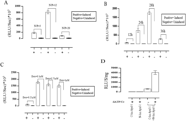
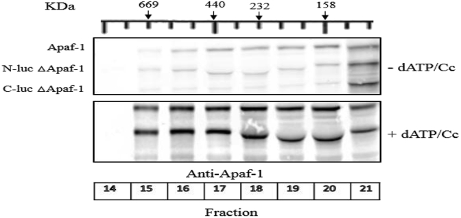
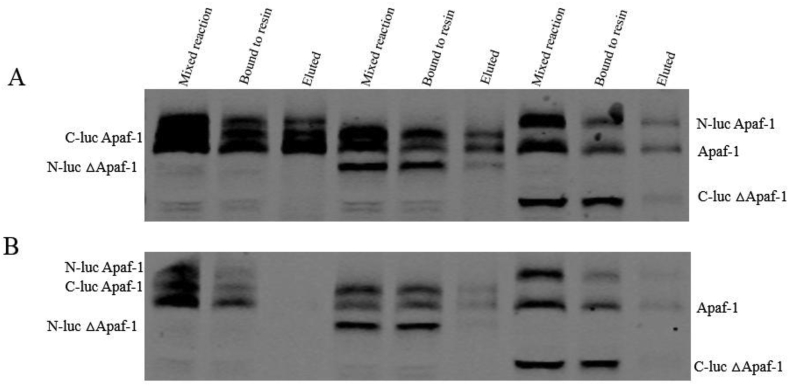
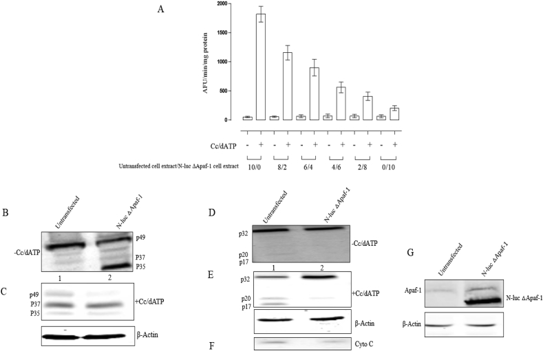
Apaf-1 is a cytosolic multi-domain protein in the apoptosis regulatory network. When cytochrome c releases from mitochondria; it binds to WD-40 repeats of Apaf-1 molecule and induces oligomerization of Apaf-1. Here in, a split luciferase assay was used to compare apoptosome formation in cell-free and cell-based systems. This assay uses Apaf-1 tagged with either N-terminal fragment or C-terminal fragment of P. pyralis luciferase. In cell based-system, the apoptosome formation is induced inside the cells which express Apaf-1 tagged with complementary fragments of luciferase while in cell-free system, the apoptosome formation is induced in extracts of the cells. In cell-free system, cytochrome c dependent luciferase activity was observed with full length Apaf-1. However, luciferase activity due to apoptosome formation was much higher in cell based system compared to cell-free system. The truncated Apaf-1 which lacks WD-40 repeats (ΔApaf-1) interacted with endogenous Apaf-1 in a different fashion compared to native form as confirmed by different retention time of eluate in gel filtration and binding to affinity column. The interactions between endogenous Apaf-1 and ΔApaf-1 is stronger than its interaction with native exogenous Apaf-1 as indicated by dominant negative effect of ΔApaf-1 on caspase-3 processing.
Keywords: Apaf-1; Apoptosis; Apoptosome; Cell death; Cell-free system; Split-luciferase; Truncated Apaf-1.
Copyright © 2018 The Authors. Published by Elsevier Inc. All rights reserved.
Figures
Similar articles
-
Loss of WD2 subdomain of Apaf-1 forms an apoptosome structure which blocks activation of caspase-3 and caspase-9.Biochimie. 2021 Jan;180:23-29. doi: 10.1016/j.biochi.2020.10.013. Epub 2020 Oct 24. Biochimie. 2021. PMID: 33132160
-
Oscillation of apoptosome formation through assembly of truncated Apaf-1.Eur J Pharmacol. 2015 Aug 5;760:64-71. doi: 10.1016/j.ejphar.2015.04.008. Epub 2015 Apr 18. Eur J Pharmacol. 2015. PMID: 25895636
-
Optimization of Experimental Variables Influencing Apoptosome Biosensor in HEK293T Cells.Sensors (Basel). 2020 Mar 23;20(6):1782. doi: 10.3390/s20061782. Sensors (Basel). 2020. PMID: 32210205 Free PMC article.
-
Apaf-1: Regulation and function in cell death.Biochimie. 2017 Apr;135:111-125. doi: 10.1016/j.biochi.2017.02.001. Epub 2017 Feb 9. Biochimie. 2017. PMID: 28192157 Review.
-
A molecular view on signal transduction by the apoptosome.Cell Signal. 2012 Jul;24(7):1420-5. doi: 10.1016/j.cellsig.2012.03.007. Epub 2012 Mar 13. Cell Signal. 2012. PMID: 22446004 Review.
Cited by
-
Aptamer-based colorimetric determination of early-stage apoptotic cells via the release of cytochrome c from mitochondria and by exploiting silver/platinum alloy nanoclusters as a peroxidase mimic.Mikrochim Acta. 2019 Nov 26;186(12):845. doi: 10.1007/s00604-019-3977-5. Mikrochim Acta. 2019. PMID: 31768654
-
MiR-27b-3p Reduces the Efficacy of Propranolol in the Treatment of Infantile Hemangioma by Inhibiting the Expression of Apaf-1.Biomedicines. 2025 Apr 30;13(5):1092. doi: 10.3390/biomedicines13051092. Biomedicines. 2025. PMID: 40426921 Free PMC article.
-
Deoxyelephantopin and Its Isomer Isodeoxyelephantopin: Anti-Cancer Natural Products with Multiple Modes of Action.Molecules. 2022 Mar 24;27(7):2086. doi: 10.3390/molecules27072086. Molecules. 2022. PMID: 35408483 Free PMC article. Review.
-
Apoptosome Formation through Disruption of the K192-D616 Salt Bridge in the Apaf-1 Closed Form.ACS Omega. 2021 Aug 24;6(35):22551-22558. doi: 10.1021/acsomega.1c02274. eCollection 2021 Sep 7. ACS Omega. 2021. PMID: 34514227 Free PMC article.
-
Bioluminescent RIPoptosome Assay for FADD/RIPK1 Interaction Based on Split Luciferase Assay in a Human Neuroblastoma Cell Line SH-SY5Y.Biosensors (Basel). 2023 Feb 20;13(2):297. doi: 10.3390/bios13020297. Biosensors (Basel). 2023. PMID: 36832063 Free PMC article.
References
-
- Schafe Z.T., Kornbluth S. The apoptosome: physiological, developmental, and pathological modes of regulation. Dev. Cell. 2006;10(5):549–561. - PubMed
-
- Riedl S.J., Salvesen G.S. The apoptosome: signalling platform of cell death. Nat. Rev. Mol. Cell Biol. 2007;8(5):405–413. - PubMed
-
- Cossarizza A. Apoptosis and HIV infection: about molecules and genes. Currr. Pharm. Des. 2008;14(3):237–244. - PubMed
-
- Evan G.I., Vousden K.H. Proliferation, cell cycle and apoptosis in cancer. Nature. 2001;411(6835):342–348. - PubMed
-
- Cain K., Bratton S.B., Cohen G.M. The Apaf-1 apoptosome: a large caspase-activating complex. Biochimie. 2002;84(2–3):203–214. - PubMed
Publication types
MeSH terms
Substances
LinkOut - more resources
Full Text Sources
Other Literature Sources
Molecular Biology Databases
Research Materials
