. 2018 Feb 6;8(1):2519.
doi: 10.1038/s41598-018-20620-w.

Identification of a novel Na+-coupled Fe3+-citrate transport system, distinct from mammalian INDY, for uptake of citrate in mammalian cells

Affiliations

Identification of a novel Na+-coupled Fe3+-citrate transport system, distinct from mammalian INDY, for uptake of citrate in mammalian cells

Jiro Ogura et al. Sci Rep. .

Retraction in

Abstract

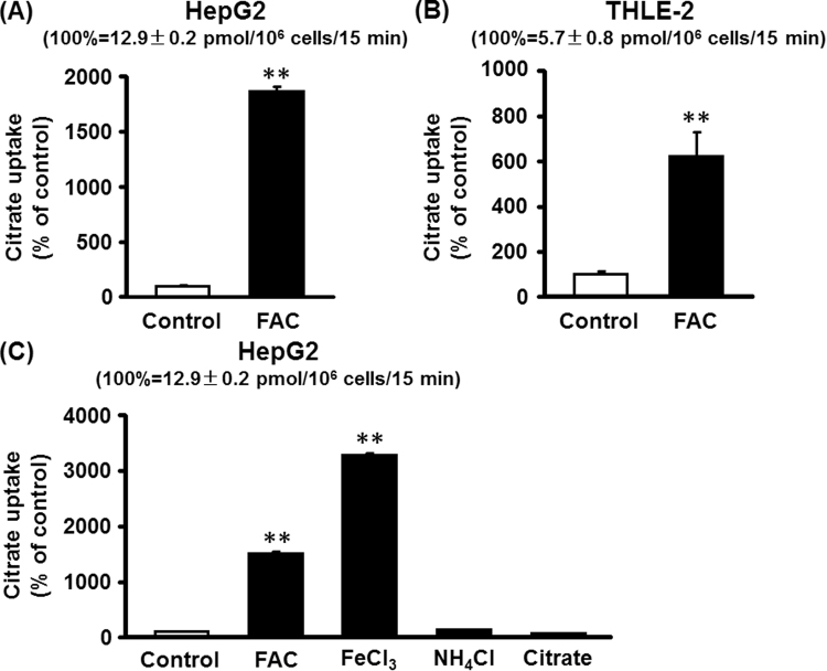
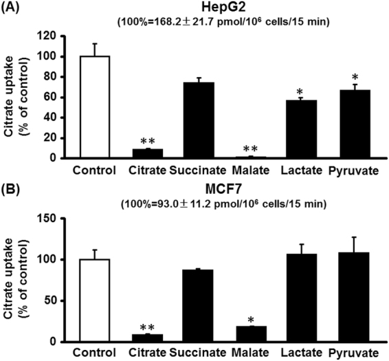
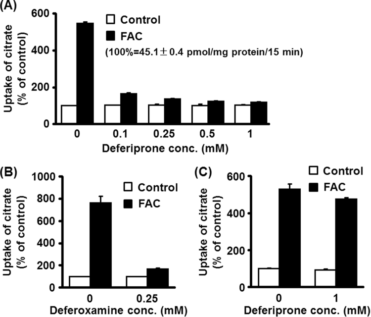
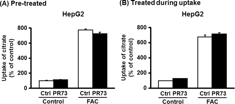
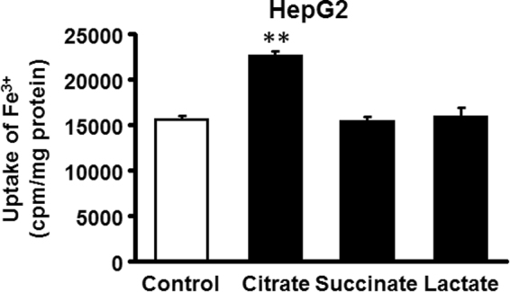
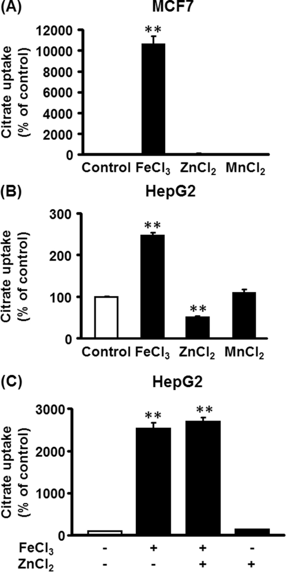
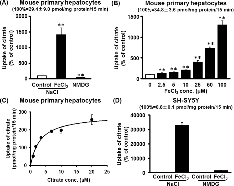
NaCT is a Na+-coupled transporter for citrate expressed in hepatocytes and neurons. It is the mammalian ortholog of INDY (I'm Not Dead Yet), a transporter which modifies lifespan in Drosophila. Here we describe a hitherto unknown transport system for citrate in mammalian cells. When liver and mammary epithelial cells were pretreated with the iron supplement ferric ammonium citrate (FAC), uptake of citrate increased >10-fold. Iron chelators abrogated the stimulation of citrate uptake in FAC-treated cells. The iron exporter ferroportin had no role in this process. The stimulation of citrate uptake also occurred when Fe3+ was added during uptake without pretreatment. Similarly, uptake of Fe3+ was enhanced by citrate. The Fe3+-citrate uptake was coupled to Na+. This transport system was detectable in primary hepatocytes and neuronal cell lines. The functional features of this citrate transport system distinguish it from NaCT. Loss-of-function mutations in NaCT cause early-onset epilepsy and encephalopathy; the newly discovered Na+-coupled Fe3+-citrate transport system might offer a novel treatment strategy for these patients to deliver citrate into affected neurons independent of NaCT. It also has implications to iron-overload conditions where circulating free iron increases, which would stimulate cellular uptake of citrate and consequently affect multiple metabolic pathways.


Conflict of interest statement

The authors declare that they have no competing interests.

Figures

Figure 1
Figure 1
Effect of pretreatment with Fe3+ on citrate uptake in a human hepatocarcinoma cell line and a human normal hepatocyte cell line. The human hepatocarcinoma cell line HepG2 (A) and the human normal hepatocyte cell line THLE-2 (B) were cultured in the absence or presence of FAC (65 μg/ml) for two passages. The cells were then seeded for uptake measurements and cultured in the absence or presence of FAC; confluent cells were used for [14C]-citrate (3.5 μM) uptake (NaCl buffer, pH 7.5; 15 min incubation). (C) HepG2 cells were cultured in the absence or presence of FAC (250 μg/ml), FeCl3 (1 mM), NH4Cl (1 mM) or citrate (1 mM) for two passages. The cells were then seeded for uptake measurements and cultured in the absence or presence of FAC, FeCl3, NH4Cl or citrate. Confluent cells were used for [14C]-citrate (3.5 μM) uptake (NaCl buffer, pH 7.5; 15 min incubation). **p < 0.01.
Figure 2
Figure 2
Non-involvement of NaCT in citrate uptake stimulated by FAC pretreatment. (A) HepG2 cells were cultured in the absence or presence of FAC (250 μg/ml) for two passages. The cells were then seeded for uptake measurements and cultured in the absence or presence of FAC; confluent cells were used for [14C]-citrate (3.5 μM) uptake (NaCl buffer, pH 7.5; 15 min incubation) in the absence or presence of 10 mM LiCl. (B) RT-PCR for expression of NaCT mRNA in control and FAC-treated HepG2 and MCF7 cells. The PCR products from both cell lines were run on the same gel to detect NaCT or HPRT (The images of the entire gels are given in Supplementary Fig. S7). (C) MCF7 cells were cultured in the absence or presence of FAC (250 μg/ml) for two passages. The cells were then seeded for uptake measurements and cultured in the absence or presence of FAC; confluent cells were used for [14C]-citrate (3.5 μM) uptake (NaCl buffer, pH 7.5; 15 min incubation) in the absence or presence of 10 mM LiCl. **p < 0.01.
Figure 3
Figure 3
Fe3+ dose-response for stimulation for citrate uptake and the Na+-dependence of the uptake process in HepG2 cells and MCF7 cells. HepG2 (A) and MCF7 cells (B) were cultured to confluence and then used for measurements of [14C]-citrate (3.5 μM) uptake (NaCl buffer, pH 7.5; 15 min incubation) in the presence of increasing concentrations of Fe3+ (FeCl3). HepG2 (C) and MCF7 cells (D) were cultured to confluence and then used for measurement of Fe3+ (50 μM)-stimulated citrate uptake in the presence (NaCl buffer, pH 7.5) or absence of Na+ (NMDG chloride, pH 7.5) using [14C]-citrate (3.5 μM). Data are presented as percent of control uptake measured in the presence of Na+ but in the absence of Fe3+. *p < 0.05; **p < 0.01.
Figure 4
Figure 4
Substrate selectivity of the citrate transport system that is stimulated in HepG2 cells and MCF7 cells by Fe3+. HepG2 (A) and MCF7 cells (B) were cultured to confluence prior to uptake measurements. Uptake of [14C]-citrate (3.5 μM) was measured in NaCl buffer, pH 7.5, for 15 min in the presence of FeCl3 (50 μM) in absence or presence of the various carboxylates (2.5 mM). *p < 0.05; **p < 0.01.
Figure 5
Figure 5
Effects of iron chelators on Fe3+-stimulated citrate uptake in FAC-treated HepG2 cells. HepG2 cells were cultured in the absence or presence of FAC (250 μg/ml) for two passages. The cells were then seeded in 24-well plates and cultured in the absence or presence of FAC (250 μg/ml) to confluence prior to uptake measurement. Uptake of [14C]-citrate (3.5 μM) was measured in a NaCl buffer, pH 7.5 for 15 min in the absence or presence of increasing concentrations of deferiprone, a cell-permeable iron chelator (A) or 0.25 mM deferoxamine, a cell-impermeable iron chelator (B). (C) HepG2 cells were cultured in the absence or presence of FAC (250 μg/ml) for two passages. The cells were then seeded in 24-well plates and cultured in the absence or presence of FAC (250 μg/ml) to confluence and then exposed to 1 mM deferoxamine prior to uptake measurement for 30 min. Uptake of [14C]-citrate (3.5 μM) was then measured in these cells in a NaCl buffer, pH 7.5 for 15 min in the absence of the iron chelator.
Figure 6
Figure 6
Influence of hepcidin mimetic minipeptide (PR73) on citrate uptake in FAC-treated HepG2 cells. HepG2 cells were cultured in the absence or presence of FAC (250 μg/ml) for two passages. (A) Prior to seeding of the cells for uptake measurements, the cells were treated with or without PR73 (1 μM) for 24 h. The cells were then prepared for uptake measurements. Uptake of [14C]-citrate (3.5 μM) was measured in a NaCl buffer, pH 7.5 for 15 min. (B) Control and FAC-treated cells were also used citrate uptake without pretreatment in the presence of PR73, but uptake was measured with PR73 present in the uptake medium during the measurement of citrate uptake.
Figure 7
Figure 7
Stimulation of Fe3+ uptake by citrate in HepG2 cells. Uptake of [59Fe]-FeCl3 was measured in HepG2 cells in NaCl buffer, pH 7.5 (15 min incubation) in the absence or presence of 50 μM citrate, succinate or lactate. The results are expressed as counts/min/mg of protein. **p < 0.01.
Figure 8
Figure 8
Effect of Zn2+ and Mn2+ on citrate uptake in MCF7 and HepG2 cells in the presence of Na+. MCF7 cells (A) and HepG2 cells (B) were cultured to confluence and the cells were used to measure the uptake [14C]-citrate (3.5 μM) for 15 min in the presence (NaCl buffer, pH 7.5) with FeCl3 (50 μM), ZnCl2 (50 μM), or MnCl2 (50 μM). (C) HepG2 cells were cultured to confluence and the cells were then used to measure the uptake of [14C]-citrate (3.5 μM) for 15 min in the presence (NaCl buffer, pH 7.5) and in the presence or absence of FeCl3 (50 μM) or ZnCl2 (50 μM) as indicated. The results are given as percent of control uptake measured in the absence of FeCl3. **p < 0.01.
Figure 9
Figure 9
Expression of the Na+-coupled Fe3+-citrate uptake system in mouse primary hepatocytes and in a human neuronal cell line. (A) Hepatocytes were cultured to confluence and then used for measurement of [14C]-citrate (3.5 μM) uptake in the presence (NaCl buffer, pH 7.5) with and without FeCl3 (50 μM) or in the absence of Na+ (NMDG chloride, pH 7.5). Data are presented as percent of control uptake measured in the presence of Na+ but in the absence of Fe3+. **p < 0.01. (B) Dose-response for Fe3+ to stimulate [14C]-citrate (3.5 μM) uptake in the presence (NaCl buffer, pH 7.5) in hepatocytes.**p < 0.01. (C) Substrate saturation kinetics for the Na+-coupled Fe3+-citrate uptake in hepatocytes. (D) The human neuronal cell line SH-SY5Y was cultured to confluence and then used for measurement of [14C]-citrate (3.5 μM) uptake in the presence (NaCl buffer, pH 7.5) with and without FeCl3 (50 μM) or in the absence of Na+ (NMDG chloride, pH 7.5). Data are presented as percent of control uptake measured in the presence of Na+ but in the absence of Fe3+.

References

    1. Bergeron MJ, Clemencon B, Hediger MA, Markovich D. SLC13 family of Na+-coupled di- and tri-carboxylate/sulfate transporters. Mol. Aspects Med. 2013;34:299–312. doi: 10.1016/j.mam.2012.12.001. - DOI - PubMed
    1. Pajor AM. Sodium-coupled dicarboxylate and citrate transporters from the SLC13 family. Pflugers Arch. 2014;466:119–130. doi: 10.1007/s00424-013-1369-y. - DOI - PubMed
    1. Rogina B, Reenan RA, Nilsen SP, Helfand SL. Extended lifespan conferred by cotransporter gene mutations in Drosophila. Science. 2000;290:2137–2140. doi: 10.1126/science.290.5499.2137. - DOI - PubMed
    1. Inoue K, Zhuang L, Maddox DM, Smith SB, Ganapathy V. Structure, function, and expression pattern of a novel sodium-coupled citrate transporter (NaCT) cloned from mammalian brain. J. Biol. Chem. 2002;277:39469–39476. doi: 10.1074/jbc.M207072200. - DOI - PubMed
    1. Rogers RP, Rogina B. The role of INDY in metabolism, health and longevity. Front. Genet. 2015;6:204. doi: 10.3389/fgene.2015.00204. - DOI - PMC - PubMed

Publication types