Review
. 2018 Jan 23:9:6.
doi: 10.3389/fphar.2018.00006. eCollection 2018.

Three-Dimensional in Vitro Cell Culture Models in Drug Discovery and Drug Repositioning

Affiliations
Review

Three-Dimensional in Vitro Cell Culture Models in Drug Discovery and Drug Repositioning

Sigrid A Langhans. Front Pharmacol. .

Abstract

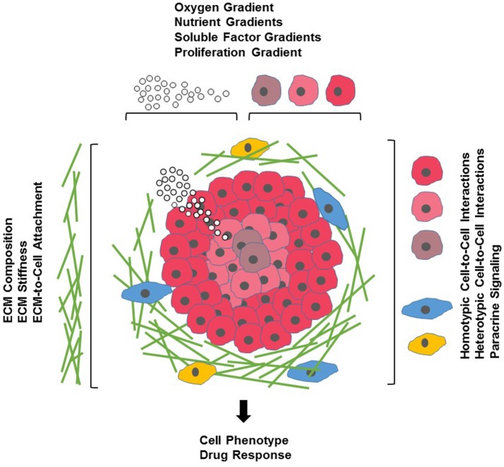
Drug development is a lengthy and costly process that proceeds through several stages from target identification to lead discovery and optimization, preclinical validation and clinical trials culminating in approval for clinical use. An important step in this process is high-throughput screening (HTS) of small compound libraries for lead identification. Currently, the majority of cell-based HTS is being carried out on cultured cells propagated in two-dimensions (2D) on plastic surfaces optimized for tissue culture. At the same time, compelling evidence suggests that cells cultured in these non-physiological conditions are not representative of cells residing in the complex microenvironment of a tissue. This discrepancy is thought to be a significant contributor to the high failure rate in drug discovery, where only a low percentage of drugs investigated ever make it through the gamut of testing and approval to the market. Thus, three-dimensional (3D) cell culture technologies that more closely resemble in vivo cell environments are now being pursued with intensity as they are expected to accommodate better precision in drug discovery. Here we will review common approaches to 3D culture, discuss the significance of 3D cultures in drug resistance and drug repositioning and address some of the challenges of applying 3D cell cultures to high-throughput drug discovery.

Keywords: extracellular matrix; high-throughput screening; hydrogel; spheroid; three-dimensional cell culture.


Figures

Figure 1
Figure 1
Cells and their microenvironment. Tissue-specific cells (red) encounter a complex microenvironment consisting of extracellular matrix (ECM) proteins and glycoproteins (green), support cells that mediate cell-cell interactions (blue), immune cells (yellow), and soluble factors (white spheres). The tissue microenvironment is further defined by physical factors such as ECM stiffness (indicated by increasing density of ECM proteins), and oxygen (indicated by red shading of tissue-specific cells) and nutrient and growth factor gradients (indicated by density of white spheres).
Figure 2
Figure 2
Types of 3D cultures. In contrast to 2D monolayers or 2.5D cultures in which cells are plated on top of a thick layer of extracellular matrix (ECM), 3D cultures form complex structures. Cells may form spheroids (anchorage independent systems) or can be encapsulated in cell culture scaffolds (anchorage dependent). Microfluidic devices and micropatterned plates with ECM components, and cultures in which formed spheroids are embedded in ECM scaffolds, can form hybrid culture systems that combine the advantages of both systems to form a complex microenvironment for 3D cell culture.

References

    1. Abaci H. E., Guo Z., Doucet Y., Jackow J., Christiano A. (2017). Next generation human skin constructs as advanced tools for drug development. Exp. Biol. Med. 242, 1657–1668. 10.1177/1535370217712690 - DOI - PMC - PubMed
    1. Ahmed T. A., Dare E. V., Hincke M. (2008). Fibrin: a versatile scaffold for tissue engineering applications. Tissue Eng. B Rev. 14, 199–215. 10.1089/ten.teb.2007.0435 - DOI - PubMed
    1. Alcaraz J., Otero J., Jorba I., Navajas D. (2017). Bidirectional mechanobiology between cells and their local extracellular matrix probed by atomic force microscopy. Semin. Cell Dev. Biol. 73, 71–81. 10.1016/j.semcdb.2017.07.020 - DOI - PubMed
    1. Alépée N., Bahinski A., Daneshian M., De Wever B., Fritsche E., Goldberg A., et al. . (2014). State-of-the-art of 3D cultures (organs-on-a-chip) in safety testing and pathophysiology. ALTEX 31, 441–477. 10.14573/altex1406111 - DOI - PMC - PubMed
    1. Alsaab H. O., Sau S., Alzhrani R., Tatiparti K., Bhise K., Kashaw S. K., et al. . (2017). PD-1 and PD-L1 Checkpoint signaling inhibition for cancer immunotherapy: mechanism, combinations, and clinical outcome. Front. Pharmacol. 8:561. 10.3389/fphar.2017.00561 - DOI - PMC - PubMed