. 2018 Jan 23:9:8.
doi: 10.3389/fpsyg.2018.00008. eCollection 2018.

Rapid Adaptation of Night Vision

Affiliations

Rapid Adaptation of Night Vision

Adam Reeves et al. Front Psychol. .

Abstract

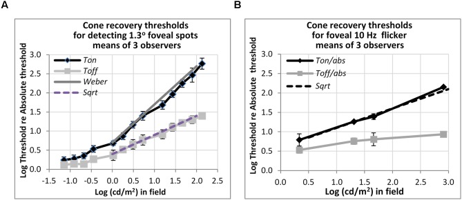
Apart from the well-known loss of color vision and of foveal acuity that characterizes human rod-mediated vision, it has also been thought that night vision is very slow (taking up to 40 min) to adapt to changes in light levels. Even cone-mediated, daylight, vision has been thought to take 2 min to recover from light adaptation. Here, we show that most, though not all adaptation is rapid, taking less than 0.6 s. Thus, monochrome (black-white-gray) images can be presented at mesopic light levels and be visible within a few 10th of a second, even if the overall light level, or level of glare (as with passing headlamps while driving), changes abruptly.

Keywords: HDR; adaptation; mesopic vision; scotopic vision; vision recovery.


Figures

FIGURE 1
FIGURE 1
(A) Schematic of the stimulus. The large circular patch containing the test disk is a uniform background field of light that defines adapting level. The four small dim spots (0.03°) guide subject’s attention to the test target location. For the rod experiments, the subjects were instructed to fixate at the fixation spot located along the edge of the background disk (5° below the test target). For the cone experiment, the subjects were fixating at the test target. (B) Timeline of the test target and background field presentations. The background field appears continuously (Ton), disappears temporarily (Toff), or flashes (Toff-flash). The test target flashed for 200 ms, as shown, or flickered for 2 s (not shown).
FIGURE 2
FIGURE 2
Plots of rod thresholds for detecting a small target (0.08°), either on a steady field (Ton) or 600 ms (Toff) or 200 ms (Toff-flash) in darkness, relative to Tabs. Thresholds were averaged over three observers and are plotted as a function of log (cd/m2) in the background field. Bars denote ± 1 standard error of the mean. Solid black line indicates the square-root prediction.
FIGURE 3
FIGURE 3
Plots of rod thresholds for detecting a larger target (1.3°), either on a steady field (Ton) or 600 ms (Toff) or 200 ms (Toff-flash) in darkness. Thresholds were averaged over three observers and are plotted as a function of log(cd/m2) in the background field. Bars denote ± 1 SE (standard error of the mean) except for Flash Off, where only +1 SE is shown. Solid black line indicates the Weber prediction and the gray line indicates the square root prediction.
FIGURE 4
FIGURE 4
Plots of relative rod thresholds, Ton/Tabs and Toff/Tabs, for perceiving 4 Hz flicker in a larger test target (1.3°). Thresholds were averaged over three observers and are plotted as a function of log(cd/m2) in the background field.
FIGURE 5
FIGURE 5
Plots of cone thresholds for (A) detecting a larger (1.3°) non-flickering target and (B) flickering target (10 Hz). Thresholds were averaged over three observers, and plotted as a function of cd/m2 in the background field. Continuous and dotted straight lines show the Weber and square-root predictions for Ton and Toff, respectively. Note that ‘abs’ here refers to the absolute threshold of the cones, not that of the rods, and corresponds (for the 1.3° test) to the dotted line shown in Figure 3.

References

    1. Aguilar M., Stiles W. S. (1954). Saturation of the rod mechanism at high levels of stimulation. Opt. Acta 1 59–65. 10.1080/713818657 - DOI
    1. Alpern M. (1961). Rhodopsin kinetics in the human eye. J. Physiol. 217 447–471. 10.1113/jphysiol.1971.sp009580 - DOI - PMC - PubMed
    1. Alpern M. (1963). Simultaneous brightness contrast for flashes of light of different durations. Invest. Ophthalmol. Vis. Sci. 2 47–54. - PubMed
    1. Baker H. D. (1961). Initial stages of dark and light adaptation. J. Opt. Soc. Am. 53 839–844. - PubMed
    1. Barlow H. B. (1957). Increment thresholds at low intensities considered as signal/noise discriminations. J. Physiol. 136 469–488. 10.1113/jphysiol.1957.sp005774 - DOI - PMC - PubMed

LinkOut - more resources