. 2018 Jan 3;5(1):171076.
doi: 10.1098/rsos.171076. eCollection 2018 Jan.

How water availability influences morphological and biomechanical properties in the one-leaf plant Monophyllaea horsfieldii

Affiliations

How water availability influences morphological and biomechanical properties in the one-leaf plant Monophyllaea horsfieldii

Tim Kampowski et al. R Soc Open Sci. .

Abstract

In its natural habitat, the one-leaf plant Monophyllaea horsfieldii (Gesneriaceae) shows striking postural changes and dramatic loss of stability in response to intermittently occurring droughts. As the morphological, anatomical and biomechanical bases of these alterations are as yet unclear, we examined the influence of varying water contents on M. horsfieldii by conducting dehydration-rehydration experiments together with various imaging techniques as well as quantitative bending and turgor pressure measurements. As long as only moderate water stress was applied, gradual reductions in hypocotyl diameters and structural bending moduli during dehydration were almost always rapidly recovered in acropetal direction upon rehydration. On an anatomical scale, M. horsfieldii hypocotyls revealed substantial water stress-induced alterations in parenchymatous tissues, whereas the cell form and structure of epidermal and vascular tissues hardly changed. In summary, the functional morphology and biomechanics of M. horsfieldii hypocotyls directly correlated with water status alterations and associated physiological parameters (i.e. turgor pressure). Moreover, M. horsfieldii showed only little passive structural-functional adaptations to dehydration in comparison with poikilohydrous Ramonda myconi.

Keywords: Monophyllaea; Ramonda; biomechanics; drought tolerance; functional morphology; turgor pressure.


Conflict of interest statement

We declare we have no competing interests.

Figures
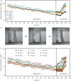
Figure 1.
Figure 1.
Changes of hypocotyl diameter in M. horsfieldii during a DRE. (a) The decrease and increase of the hypocotyl diameter at a marked position (30 mm above ground) of five different plants are shown in different colours. The majority of plants regain their original hypocotyl diameter after rehydration. (b) The three images exemplarily show the change of hypocotyl diameter in the aforementioned DRE. The hypocotyl shown belongs to plant two in (a). The time scale and scale bars are indicated. (c) The diameter changes along the hypocotyl of M. horsfieldii at 5 mm intervals are illustrated. Each colour represents one of nine different hypocotyl positions between 10 and 50 mm above ground.
Figure 2.
Figure 2.
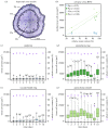
Anatomical changes in the tissues of M. horsfieldii hypocotyls during a DRE. (a) From outside to inside, the hypocotyl consists of four tissue regions: epidermis (Ep), peripheral parenchyma ring (Par), vascular bundle ring (Vbr) and central parenchyma cylinder (Pac). (b) In contrast to the cells of the epidermis and the vascular bundle ring, those of the parenchymatous tissues show a strong correlation between RWC and median cell area (Ep, blue open circles; Par, green circles; Vbr, light-blue open squares; Pac, light-green squares). Pearson's correlation coefficients (r) are given in parentheses. (c–f) The parenchymatous tissues exhibit the highest cell area changes during dehydration (days 1–9) and rehydration (days 9–10) of the hypocotyl (green and light-green boxplots), whereas those of the epidermis and vascular bundle ring hardly change at all (blue and light-blue boxplots). The RWC alteration is indicated by purple diamonds. Significant changes are marked by capital letters above individual boxplots. The corresponding sample sizes (N) are indicated below each boxplot.
Figure 3.
Figure 3.
Change in the structural bending modulus of M. horsfieldii hypocotyls during a DRE. Illustration of the changes in structural bending modulus during DRE according to the alterations of the RWC (black circles). The significant reduction of the structural bending modulus during dehydration (days 1–5) and its recovery after rehydration (days 5–6) are highlighted. The RWC alteration is indicated by purple diamonds. N, sample size; **p < 0.01; ***p < 0.001.
Figure 4.
Figure 4.
Relationship between turgor pressures and RWCs in different tissues of M. horsfieldii hypocotyls. In the cells of the epidermis (blue open circles), the parenchyma ring (green circles) and the parenchyma cylinder (light-green squares), the turgor pressure correlates positively with the RWC (Spearman's correlation coefficients are given). Only the parenchyma cells in the vascular bundle ring region (light-blue open squares) show no correlation between the RWC and the turgor pressure. The ρ values are given to show a general trend. However, it has to be kept in mind that for a reliable statistical analysis, much larger sample sizes would be necessary. N, sample size; ρ, correlation coefficient.
Figure 5.
Figure 5.
Comparison of the regeneration capacities of M. horsfieldii hypocotyl and R. myconi leaf stalk segments after different drying durations. (a,b) The regeneration capability of M. horsfieldii hypocotyl segments is considerably less than that of R. myconi leaf stalk segments after different drying durations. (c) Comparisons of M. horsfieldii and R. myconi regeneration capacities after different drying durations indicate different amounts of passively functioning adaptations. Values are given as means ± standard errors; N = 10; *p < 0.05; **p < 0.01; ***p < 0.001; DW, dry weight; FW, fresh weight; RW, rehydrated weight.

References

    1. Kiew R. 2009. The natural history of Malaysian Gesneriaceae. Malay. Nat. J. 61, 257–265.
    1. Green TGA, Sancho LG, Pintado A. 2011. Ecophysiology of desiccation/rehydration cycles in mosses and lichens. In Plant desiccation tolerance (eds Lüttge U, Beck E, Bartels D), pp. 89–120. Berlin, Germany: Springer.
    1. Chin SC. 1977. The limestone hill flora of Malaya I. Gard. Bull. Singap. 30, 165–219.
    1. Figdor W. 1903. Über Regeneration bei Monophyllaea horsfieldii R.Br. Österr. Bot. Z. 53, 393–396. (doi:10.1007/BF01791210) - DOI
    1. Ayano M, Imaichi R, Kato M. 2005. Developmental morphology of the Asian one-leaf plant, Monophyllaea glabra (Gesneriaceae) with emphasis on inflorescence morphology. J. Plant Res. 118, 99–109. (doi:10.1007/s10265-005-0195-5) - DOI - PubMed

LinkOut - more resources