Review
. 2018 Mar;135(3):311-336.
doi: 10.1007/s00401-018-1815-1. Epub 2018 Feb 6.

Functional morphology of the blood-brain barrier in health and disease

Affiliations
Review

Functional morphology of the blood-brain barrier in health and disease

Stefan Liebner et al. Acta Neuropathol. 2018 Mar.

Abstract

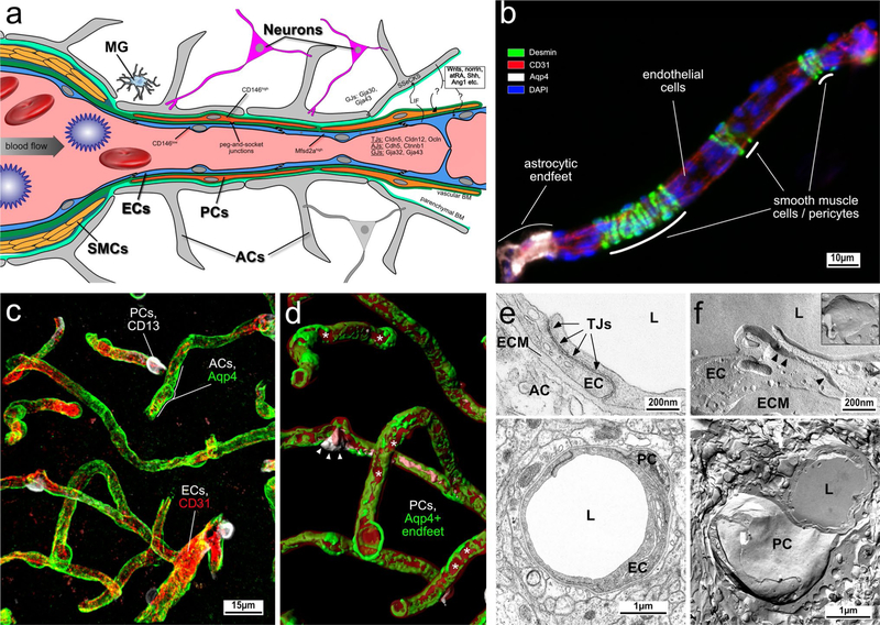
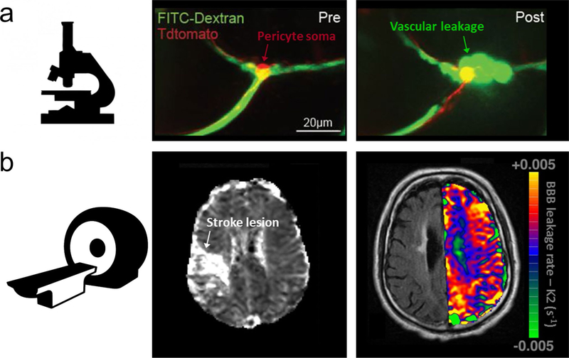
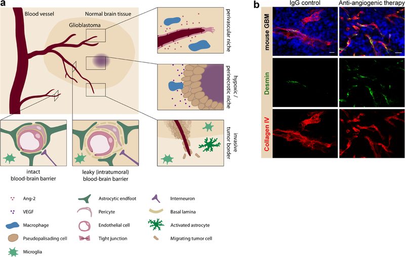
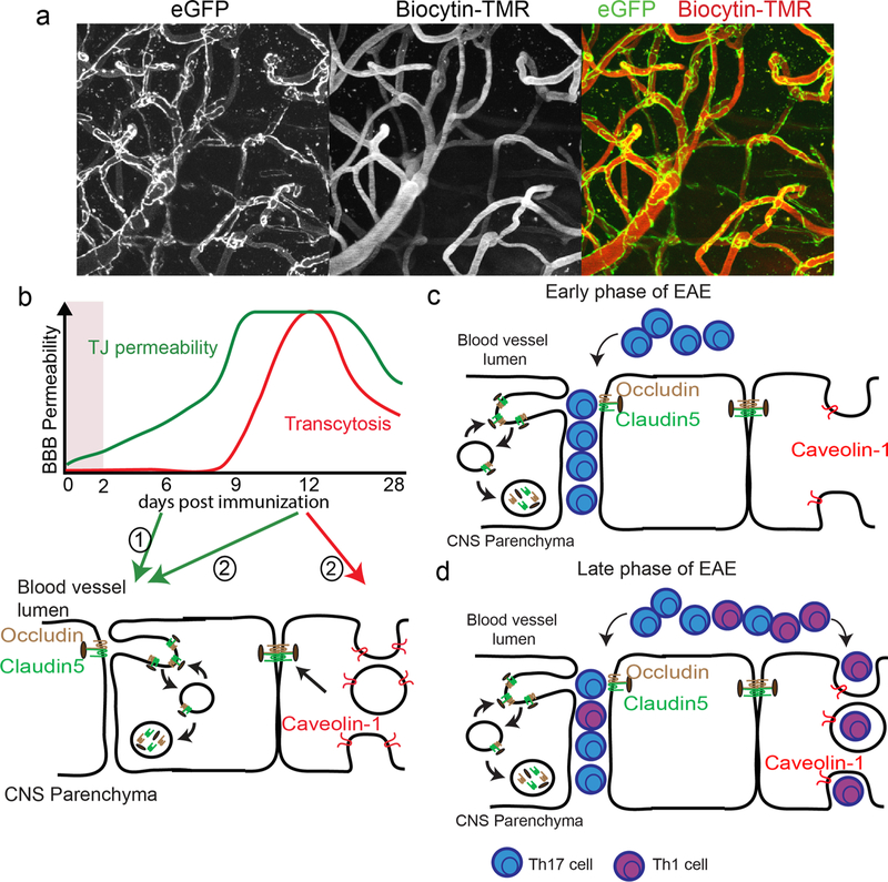
The adult quiescent blood-brain barrier (BBB), a structure organised by endothelial cells through interactions with pericytes, astrocytes, neurons and microglia in the neurovascular unit, is highly regulated but fragile at the same time. In the past decade, there has been considerable progress in understanding not only the molecular pathways involved in BBB development, but also BBB breakdown in neurological diseases. Specifically, the Wnt/β-catenin, retinoic acid and sonic hedgehog pathways moved into the focus of BBB research. Moreover, angiopoietin/Tie2 signalling that is linked to angiogenic processes has gained attention in the BBB field. Blood vessels play an essential role in initiation and progression of many diseases, including inflammation outside the central nervous system (CNS). Therefore, the potential influence of CNS blood vessels in neurological diseases associated with BBB alterations or neuroinflammation has become a major focus of current research to understand their contribution to pathogenesis. Moreover, the BBB remains a major obstacle to pharmaceutical intervention in the CNS. The complications may either be expressed by inadequate therapeutic delivery like in brain tumours, or by poor delivery of the drug across the BBB and ineffective bioavailability. In this review, we initially describe the cellular and molecular components that contribute to the steady state of the healthy BBB. We then discuss BBB alterations in ischaemic stroke, primary and metastatic brain tumour, chronic inflammation and Alzheimer's disease. Throughout the review, we highlight common mechanisms of BBB abnormalities among these diseases, in particular the contribution of neuroinflammation to BBB dysfunction and disease progression, and emphasise unique aspects of BBB alteration in certain diseases such as brain tumours. Moreover, this review highlights novel strategies to monitor BBB function by non-invasive imaging techniques focussing on ischaemic stroke, as well as novel ways to modulate BBB permeability and function to promote treatment of brain tumours, inflammation and Alzheimer's disease. In conclusion, a deep understanding of signals that maintain the healthy BBB and promote fluctuations in BBB permeability in disease states will be key to elucidate disease mechanisms and to identify potential targets for diagnostics and therapeutic modulation of the BBB.

Keywords: Alzheimer’s disease; Blood–brain barrier; Brain tumour; Neuroinflammation; Steady state; Stroke.


Conflict of interest statement

Conflict of interest The authors declare that they have no conflict of interest.

Figures

Fig. 1
Fig. 1
Morphological and functional characteristics of the healthy NVU and BBB. a Scheme of brain vessels ranging from an arteriole, via a capillary to a venule, representing the cellular and molecular composition of the NVU. b Isolated mouse cortical micro vessel stained for CD31, desmin, Aqp4 and DAPI. Due to the mechanical and chemical isolation process, only remnants of PCs and AC end-feet are detectable at the CD31 + vessel. c Mouse cortical vessels stained in vibratome sections for CD31, CD13 and Aqp4, demonstrating almost complete coverage of vessels by AC endfeet. PCs indicated by arrowheads are located on microvessels and cling around them with their cellular processes. d Magnified ROI of C and 3D rendered, demonstrating partially incomplete astrocytic end-feet coverage of endothelial cells (asterisks) as well as PC cell body (arrowheads) and processes (arrows). e Transmission electron microscopy showing a cortical capillary with ECs, PCs and ACs with interendothelial junctions (lower panel). Higher magnification of interendothelial junctions (upper panel) (kindly provided by Jadranca Macas, Institute of Neurology, Goethe University Clinic Frankfurt, Germany). f Freeze fracture preparation of a cortical capillary with an attached pericyte (lower panel). Freeze fracture of interendothelial junction (upper panel), arrowheads show junctional strands. Inset shows junction strands in higher magnification (kindly provided by Hartwig Wolburg, Institute of Pathology, University Clinic Tübingen, Germany). ECs, endothelial cells; PCs, pericytes; SMCs, smooth muscle cells; ACs, astrocytes; MG, microglia
Fig. 2
Fig. 2
Imaging BBB permeability after stroke—from microscopic detection in mice to whole-brain assessment in patients. a Two-photon microscopic imaging, pre and post photothrombotic vessel occlusion, showing post-stroke capillary BBB leakage (green arrow) occurring particularly at pericyte somata (red arrow) in mouse brain (kindly provided by Robert Underly and Andy Shih, Medical University of South Carolina, Charleston, SC, USA) (for details see [127]). b MRI of increased BBB leakage rate (right image; colour coding) in contralesional white matter in a patient with an acute unilateral ischaemic stroke lesion (left image; hyperintensity on perfusion MRI-derived mean transit time map) (kindly provided by Ona Wu, Massa-chusetts General Hospital and Harvard Medical School, Boston, MA, USA) (Reprinted by Permission of SAGE Publications, Ltd.) (for details see [104])
Fig. 3
Fig. 3
Schematic representation of the glioblastoma microenvironment. a Glioblastoma defining niches are displayed. Perivascular niche: Glioma and tumour stroma cells create a specialised vascular niche. Macrophages are the most abundant population that support the tumour growth by releasing proangiogenic factors. Hypoxic/perinecrotic niche: Necrotic areas are characterised by pseudopalisading cells and high levels of hypoxia which recruit tumour supporting macrophages. Invasive tumour border: Glioma cells are traversing the blood vessels. Peripheral blood vessels preserve intact blood–brain properties in contrast to the tumour vasculature which is leaky. b Combining anti-VEGF therapy with Ang-2 blockade acts synergistically on vascular normalisation. Immunofluorescence staining with antibodies directed against Collagen IV (red) and desmin (green) on vibratome sections of GL261 glioma bearing mice, untreated and after dual anti-VEGF (Aflibercept) and Ang-2 inhibition (Trebananib)
Fig. 4
Fig. 4
Schematic presentation of BBB breakdown and lymphocyte trafficking across a damaged BBB in neuroinflammation. a Transgenic mice that allow visualisation of endothelial tight junctions (Tg eGFP::Claudin-5) with intravital two-photon microscopy. The image shows cortical blood vessels that have eGFP in tight junctions (green). Biocytin-TMR tracer (red) was injected into the tail vein of mice to visualise the vascular tree. This tracer also allows measurement of BBB permeability. b A working model of BBB disruption during EAE. Increased paracellular BBB permeability (green line) occurs prior to onset and persists throughout EAE. Transcellular BBB permeability (red line) only transiently elevates at acute disease. Para-cellular permeability is due to rapid remodelling of TJ proteins, while transcellular permeability results from enhanced caveolar trafficking. c During the early phase of MS/EAE progression, there is an initial breakdown of endothelial cells TJs at the BBB that causes an increase in paracellular permeability. This allows the preferential entry of Th17 lymphocytes (blue) in the early phase of the disease, although some Th1 lymphocytes (purple) also enter through this route. b The increase in caveolar transport (transcellular permeability) through upregulation of Caveolin-1 is observed only during the late phase of neuroinflammation at the peak of the disease. Th1 lymphocytes (purple) preferentially use caveolae to cross the BBB through enhance transcellular permeability. TJ remodelling in both phases of the disease involves formation of membrane invaginations (protrusions) that fuse with EEA1+ endosomes. However, TJ remodelling is independent of caveolae suggesting that in MS/EAE, similar to BBB development, there are distinct mechanisms that impair paracellular versus transcellular barrier properties of the CNS vasculature
Fig. 5
Fig. 5
Schematic representation of BBB alterations and leukocyte extravasation in postcapillary venules in the AD brain. Endothelial cells are the first barrier between blood leukocytes and the brain parenchyma. Endothelial cells are linked by tight junctions closely surrounded by pericytes and encircled by the endothelial basal lamina (brown line) and parenchymal basement membrane (violet line). Astrocyte endfeet processes support endothelial functions and provide the cellular link to neuronal cells. Aβ and other inflammatory stimuli promote the activation of vascular endothelium, potentially inducing the expression of adhesion molecules and chemoattractants. The activated endothelium promotes the adhesion of neutrophils and eventually other circulating leukocytes that transmigrate into the brain parenchyma. Neutrophils adhered on the activated endothelium may release neutrophil extracellular traps (NETs) comprising decondensed chromatin and active proteases, which damage the BBB. Migrated neutrophils release inflammatory mediators and NETs, and may damage neurons. Neutrophils and glial cells may become trapped in a cycle of reciprocal activation, promoting chronic inflammation and neurodegeneration

Similar articles

Cited by

References

    1. Abadier M, Haghayegh Jahromi N, Cardoso Alves L, Boscacci R, Vestweber D, Barnum S, Deutsch U, Engelhardt B, Lyck R (2015) Cell surface levels of endothelial ICAM-1 influence the transcellular or paracellular T-cell diapedesis across the blood–brain barrier. Eur J Immunol 45:1043–1058. 10.1002/eji.201445125 - DOI - PubMed
    1. Abulrob A, Brunette E, Slinn J, Baumann E, Stanimirovic D (2008) Dynamic analysis of the blood–brain barrier disruption in experimental stroke using time domain in vivo fluorescence imaging. Mol Imaging 7:248–262. 10.2310/7290.2008.00025 - DOI - PubMed
    1. Agarwal R, Brunelli SM, Williams K, Mitchell MD, Feldman HI, Umscheid CA (2009) Gadolinium-based contrast agents and nephrogenic systemic fibrosis: a systematic review and meta-analysis. Nephrol Dial Transplant 24:856–863. 10.1093/ndt/gfn593 - DOI - PubMed
    1. Allen E, Jabouille A, Rivera LB, Lodewijckx I, Missiaen R, Steri V, Feyen K, Tawney J, Hanahan D, Michael IP, Bergers G (2017) Combined antiangiogenic and anti-PD-L1 therapy stimulates tumor immunity through HEV formation. Sci Transl Med 10.1126/scitranslmed.aak9679 - DOI - PMC - PubMed
    1. Alvarez JI, Dodelet-Devillers A, Kebir H, Ifergan I, Fabre PJ, Terouz S, Sabbagh M, Wosik K, Bourbonnière L, Bernard M, van Horssen J, de Vries HE, Charron F, Prat A (2011) The hedgehog pathway promotes blood–brain barrier integrity and CNS immune quiescence. Science 334:1727–7731. 10.1126/science.1206936 - DOI - PubMed

Publication types

MeSH terms

LinkOut - more resources