

The Role of Tendon Transfers for Irreparable Rotator Cuff Tears
- PMID: 29411320
- PMCID: PMC5825349
- DOI: 10.1007/s12178-018-9468-1
The Role of Tendon Transfers for Irreparable Rotator Cuff Tears
Abstract
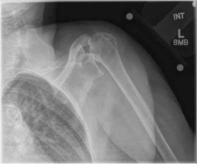
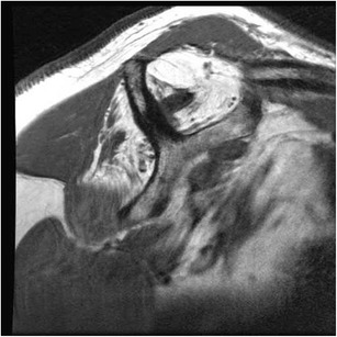
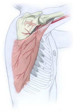
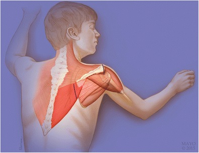
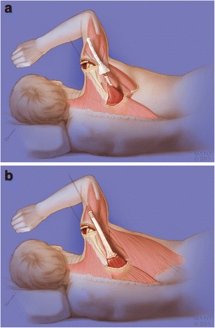
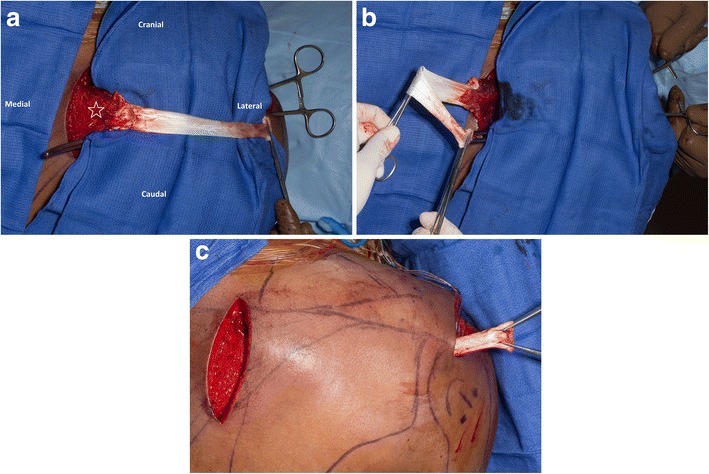
Purpose of review: This review aims to describe the tendon transfer options for treating irreparable rotator cuff tears (RCTs). Options for transfer include latissimus dorsi and lower trapezius transfers for posterior-superior RCTs and pectoralis major and latissimus dorsi transfer for anterior-superior RCTs.
Recent findings: While the latissimus dorsi tendon transfer has historically been performed for posterosuperior RCTs, the lower trapezius transfer is a more anatomic option and has demonstrated promising results in recent studies. Similarly, the pectoralis major transfer has historically been the tendon transfer of choice for anterosuperior RCTs. However, the latissimus dorsi tendon transfer has recently been shown to be a safe and anatomic tendon transfer for subscapularis insufficiency. The treatment of irreparable RCTs involves complex decision making. Tendon transfer procedures can restore the glenohumeral joint force couples, allowing restoration of near-normal shoulder kinematics. Benefits include reliable pain relief, increased function, and increased strength. Proper selection of donor tendon is crucial, and the principles of tendon transfer procedures must be adhered to for maximal benefit.
Keywords: Latissimus dorsi; Lower trapezius; Pectoralis major; Rotator cuff tear; Tendon transfer.
Conflict of interest statement
Conflict of Interests
The authors whose names are listed above certify that they have NO affiliations with or involvement in any organization or entity with any financial interest (such as honoraria; educational grants; participation in speakers’ bureaus; membership, employment, consultancies, stock ownership, or other equity interest; and expert testimony or patent-licensing arrangements), or non-financial interest (such as personal or professional relationships, affiliations, knowledge, or beliefs) in the subject matter or materials discussed in this manuscript.
Human and Animal Rights and Informed Consent
This article does not contain any studies with human or animal subjects performed by any of the authors.
Figures
References
-
- Burkhart SS. Arthroscopic treatment of massive rotator cuff tears. Clinical results and biomechanical rationale. Clin Orthop Relat Res. 1991;267:45–56. - PubMed
-
- Goutallier D, Postel JM, Bernageau J, Lavau L, Voisin MC. Fatty muscle degeneration in cuff ruptures. Pre- and postoperative evaluation by CT scan. Clin Orthop Relat Res. 1994;304:78–83. - PubMed
Publication types
LinkOut - more resources
Full Text Sources
Other Literature Sources
Research Materials
