. 2018 May;79(5):2451-2459.
doi: 10.1002/mrm.27122. Epub 2018 Feb 6.

Probing cardiac metabolism by hyperpolarized 13C MR using an exclusively endogenous substrate mixture and photo-induced nonpersistent radicals

Affiliations

Probing cardiac metabolism by hyperpolarized 13C MR using an exclusively endogenous substrate mixture and photo-induced nonpersistent radicals

Jessica A M Bastiaansen et al. Magn Reson Med. 2018 May.

Abstract

Purpose: To probe the cardiac metabolism of carbohydrates and short chain fatty acids simultaneously in vivo following the injection of a hyperpolarized 13 C-labeled substrate mixture prepared using photo-induced nonpersistent radicals.

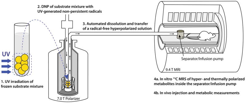
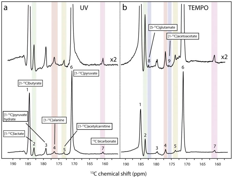
Methods: Droplets of mixed [1-13 C]pyruvic and [1-13 C]butyric acids were frozen into glassy beads in liquid nitrogen. Ethanol addition was investigated as a means to increase the polarization level. The beads were irradiated with ultraviolet light and the radical concentration was measured by ESR spectroscopy. Following dynamic nuclear polarization in a 7T polarizer, the beads were dissolved, and the radical-free hyperpolarized solution was rapidly transferred into an injection pump located inside a 9.4T scanner. The hyperpolarized solution was injected in healthy rats to measure cardiac metabolism in vivo.

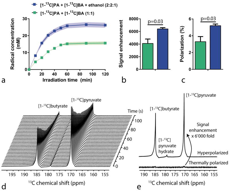
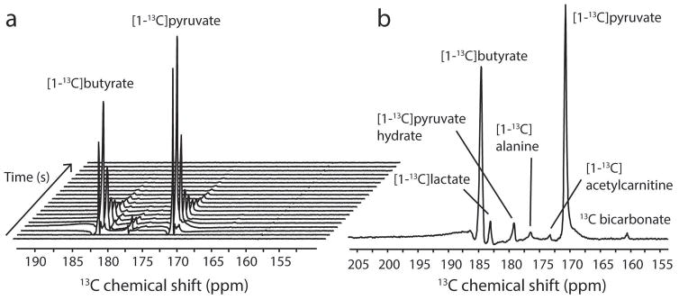
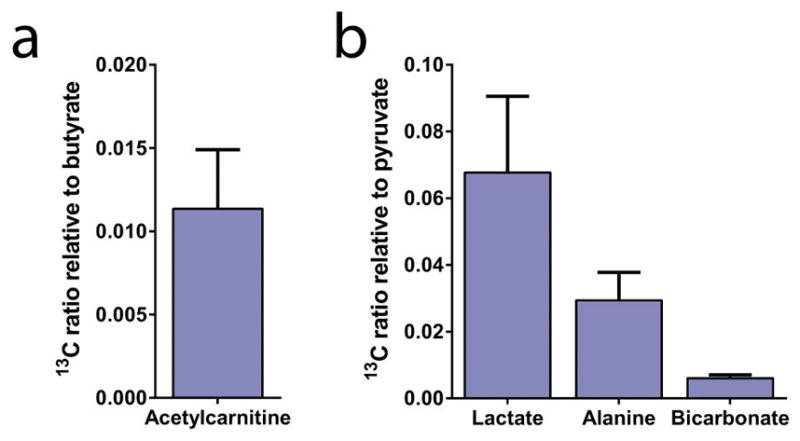
Results: Ultraviolet irradiation created nonpersistent radicals in a mixture containing 13 C-labeled pyruvic and butyric acids, and enabled the hyperpolarization of both substrates by dynamic nuclear polarization. Ethanol addition increased the radical concentration from 16 to 26 mM. Liquid-state 13 C polarization was 3% inside the pump at the time of injection, and increased to 5% by addition of ethanol to the substrate mixture prior to ultraviolet irradiation. In the rat heart, the in vivo 13 C signals from lactate, alanine, bicarbonate, and acetylcarnitine were detected following the metabolism of the injected substrate mixture.

Conclusion: Copolarization of two different 13 C-labeled substrates and the detection of their myocardial metabolism in vivo was achieved without using persistent radicals. The absence of radicals in the solution containing the hyperpolarized 13 C-substrates may simplify the translation to clinical use, as no radical filtration is required prior to injection.

Keywords: carbon-13; energy metabolism; hyperpolarization; metabolic imaging; oxidative metabolism.


Conflict of interest statement

Competing financial interests

Arnaud Comment is currently employed by General Electric Medical Systems, Inc.

Figures

Figure 1
Figure 1
Illustration of the in vivo and in vitro experimental method based on UV-irradiated 13C-labeled metabolic substrate mixtures: (1) a frozen mixture of 13C-labeled substrates, in this case [1-13C]butyric and [1-13C]pyruvic acids, is irradiated with UV light at 77 K; (2) the mixture is loaded into a DNP polarizer and 13C nuclei are dynamically polarized for 1–2 h; (3) using an automated process, the sample is quickly dissolved in superheated buffer solution and automatically transferred from the polarizer into a separator/injection pump located inside the bore of an MRI magnet; (4a & 4b) in vitro 13C MRS measurements are performed while the hyperpolarized 13C-substrate mixture is inside the separator/injection pump and/or the mixture is injected into the rat via a femoral vein catheter and in vivo hyperpolarized 13C MRS measurements are launched.
Figure 2
Figure 2
a) Radical concentration as a function of the UV light irradiation time for two different mixtures: [1-13C]pyruvic- and [1-13C]butyric acids in a 1:1 volume ratio (green), and the same with the addition of ethanol (blue). Signal intensity enhancement (b) and polarization levels (c) of pyruvic acid using two differently prepared mixtures. (d,e) In vitro hyperpolarized 13C MRS of the UV-irradiated mixture of 13C labeled substrates. (d) A hyperpolarized 13C spectral time course of the mixture measured in the infusion pump inside the MRI scanner after dissolution and automated sample transfer. Hyperpolarized spectra were acquired with an RF excitation angle of 5° and repetition time TR of 3 s. (e) The first measured hyperpolarized 13C spectrum after dissolution (top spectrum) was compared to its thermally polarized counterpart (bottom spectrum) to calculate the SNR enhancement in liquid state. A 6’000 fold signal enhancement was measured. All spectra are displayed without spectral line broadening.
Figure 3
Figure 3
In vivo 13C MRS measured in the healthy myocardium after the injection of a hyperpolarized UV-irradiated [1-13C]pyruvate/[1-13C]butyrate mixture. (A) Representative 13C spectral time course following the injection of the substrate mixture. Acquisitions were respiratory-gated and cardiac-triggered with a repetition time of 3 s and a flip angle of 30°. (B) Sum of the 15 spectra acquired starting 9 s after dissolution. The myocardial metabolism of both pyruvate and butyrate could be observed through the formation of alanine, lactate, bicarbonate and acetylcarnitine.
Figure 4
Figure 4
13C metabolite signal integral ratios relative to their respective injected substrate following the injection and myocardial metabolism of a UV-irradiated radical-free mixture of [1-13C]pyruvic acid and [1-13C]butyric acid.
Figure 5
Figure 5
Comparison of spectra acquired in vivo after the administration of a UV-irradiated mixture containing [1-13C]pyruvate and [1-13C]butyrate (left panel) and the administration of a mixture of [1-13C]pyruvate and [1-13C]butyrate containing TEMPO persistent radicals (right panel). Data obtained using TEMPO radicals was presented in an earlier publication (14). Spectra in the top panels were scaled with factor 2. Observed metabolites are indicated as follows: 1, [1-13C]butyrate; 2, [1-13C]lactate; 3, [1-13C]pyruvate hydrate; 4, [1-13C]alanine; 5, [1-13C]acetylcarnitine; 6, [1-13C]pyruvate; 7, 13C-bicarbonate; 8, [5-13C]glutamate; 9, [1-13C]acetoacetate.

References

    1. Ardenkjaer-Larsen JH, Fridlund B, Gram A, Hansson G, Hansson L, Lerche MH, Servin R, Thaning M, Golman K. Increase in signal-to-noise ratio of > 10,000 times in liquid-state NMR. Proc Natl Acad Sci U S A. 2003;100(18):10158–10163. - PMC - PubMed
    1. Golman K, in’t Zandt R, Thaning M. Real-time metabolic imaging. Proceedings of the National Academy of Sciences of the United States of America. 2006;103(30):11270–11275. - PMC - PubMed
    1. Brindle KM. Imaging Metabolism with Hyperpolarized C-13-Labeled Cell Substrates. Journal of the American Chemical Society. 2015;137(20):6418–6427. - PubMed
    1. Comment A. Dissolution DNP for in vivo preclinical studies. J Magn Reson. 2016;264:39–48. - PubMed
    1. Comment A, Merritt ME. Hyperpolarized magnetic resonance as a sensitive detector of metabolic function. Biochemistry. 2014;53(47):7333–7357. - PMC - PubMed

Publication types