Review
. 2018 Aug 15;9(8):1872-1883.
doi: 10.1021/acschemneuro.7b00456. Epub 2018 Feb 20.

Drosophila as a Model System for Neurotransmitter Measurements

Affiliations
Review

Drosophila as a Model System for Neurotransmitter Measurements

Mimi Shin et al. ACS Chem Neurosci. .

Abstract

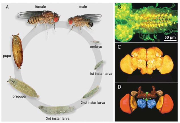
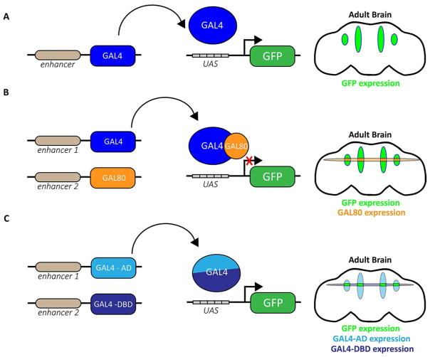
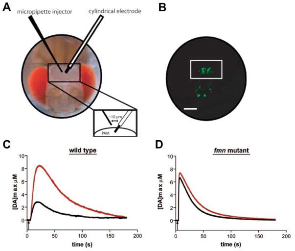
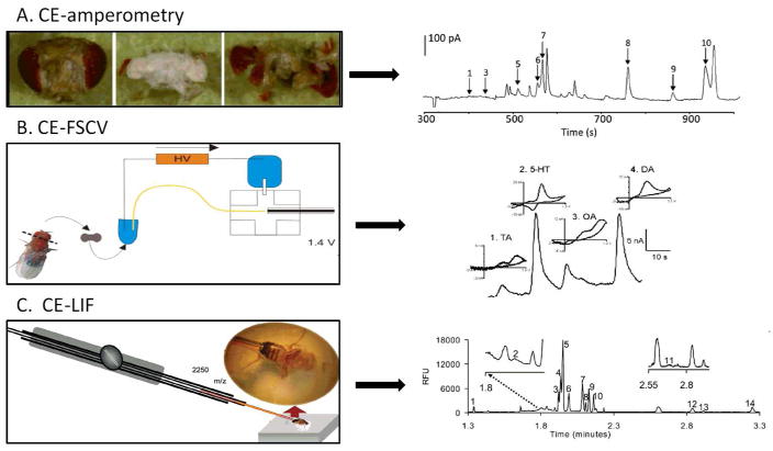
Drosophila melanogaster, the fruit fly, is an important, simple model organism for studying the effects of genetic mutations on neuronal activity and behavior. Biologists use Drosophila for neuroscience studies because of its genetic tractability, complex behaviors, well-known and simple neuroanatomy, and many orthologues to human genes. Neurochemical measurements in Drosophila are challenging due to the small size of the central nervous system. Recently, methods have been developed to measure real-time neurotransmitter release and clearance in both larvae and adults using electrochemistry. These studies have characterized dopamine, serotonin, and octopamine release in both wild type and genetic mutant flies. Tissue content measurements are also important, and separations are predominantly used. Capillary electrophoresis, with either electrochemical, laser-induced fluorescence, or mass spectrometry detection, facilitates tissue content measurements from single, isolated Drosophila brains or small samples of hemolymph. Neurochemical studies in Drosophila have revealed that flies have functioning transporters and autoreceptors, that their metabolism is different than in mammals, and that flies have regional, life stage, and sex differences in neurotransmission. Future studies will develop smaller electrodes, expand optical imaging techniques, explore physiological stimulations, and use advanced genetics to target single neuron release or study neurochemical changes in models of human diseases.

Keywords: Drosophila; capillary electrophoresis; dopamine; glutamate; octopamine; optogenetics; serotonin; voltammetry.


Figures

Figure 1
Figure 1
(A) Drosophila melanogaster develops in approximately 10 days, moving through embryo, three larval, pupal, and adult stages. (B) Ddc-GAL4 drives GFP expression (green) in dopamine and serotonin neurons of the larval brain. Antibody detecting serotonin (red) present; overlap of cells expressing GFP and serotonin appears yellow. (C) Three-dimensional imaging of the adult brain reveals a few distinct anatomical regions. (D) Anatomical regions within the brain were labeled. Dark brown: mushroom body; blue: antennal lobe; green: central complex; red: medulla; orange: lobula. Figures reprinted from ref [6] and [27], with permission of Elsevier.
Figure 2
Figure 2
(A) In flies carrying both the GAL4 (blue) and the UAS constructs, GAL4 will drive expression of the transgene. In this example, GAL4 drives expression of transgenic GFP in four regions of the adult brain. (B) GAL80 (orange) represses GAL4 transcriptional activity, and in cells expressing both GAL4 (blue) and GAL80, transgenic GFP expression will be restricted. (C) With split-GAL4, transgenic GFP (green) will only be expressed in the limited number of cells expressing both the GAL4 activating domain (GAL4-AD, light blue) and the GAL4 DNA-binding domain (GAL4-DBD, dark blue).
Figure 3
Figure 3
In vivo measurement of exogenously applied dopamine. (A) Adult fly head after microsurgery exposing PAM neurons (scale bar = 100 μm). (B) Fluorescence image of microsugeried fly head showing GPF expressed in dopaminergic neurons and white box highlights PAM neurons. (C) Representative wild type (WT) data of exogenously applied dopamine trace before (black line) after (red line) the brain was exposed to 1.0 mM cocaine. (D) Exogenously applied dopamine in fumin mutant flies, where dopamine concentration is significantly increased compared to WT. Figures modified from ref [39], with permission of American Chemical Society.
Figure 4
Figure 4
CsChrimson stimulated dopamine release from Drosophila larvae. (A) CFME in protocerebrum. Scale bar is 50 μm (top). Concentration trace of CsChrimson evoked dopamine release in the larval protocerebrum (2s continuous stimulation) with cyclic voltammogram (CV) of released dopamine (bottom). (B) CFME in larval VNC. Scale bar is 50 μm (top). Concentration trace and CV of CsChrimson evoked dopamine release in the larval VNC (2s continuous stimulation) (bottom). Figures modified from reference [51], with permission of John Wiley and Sons.
Figure 5
Figure 5
Neurochemical tissue content using different separations. (A) Image of a freeze-dried adult Drosophila head (left), extracted brain (center), and the cuticle (right). Electropherogram of freeze-dried dissected 15 brain homogenates with 10 Identified peaks (right) (1) dopamine, (2) salsolinol, (3) N-acetyloctopamine, (4) octopamine,(5) N-acetylserotonin, (6) N-acetyltyramine, (7) N-acetyldopamine, (8) L-DOPA, (9) catechol, and (10) tyramine. (B) Schematic diagram of CE-FSCV instrument (left). Electropherogram separation of tyramine, serotonin, octopamine, and dopamine in a single brain (Right). Cyclic voltammograms of each analyte are labeled at the corresponding peak. (C) Schematic diagram of a Drosophila hemolymph sampling setup (left). Electropherogram for fluorescamine labeled amino acids of wildtype adult fly hemolymph (right). (1) arginine, (2) citrulline, (3) tyrosine, (4) histidine, (5) glutamine, (6) asparagine and threonine, (7) alanine and serine, (8) taurine, (9) lysine, (10) glycine, (11) cysteine, (12) glutamate, (13) aspartate, and (14) unknown. Panel A modified from ref. [79], Panel B from [80], and Panel C from [85], all with permission from the American Chemical Society.
Figure 6
Figure 6
Nanometer scale carbon electrodes. (A) Scanning electron microscopy (SEM) image of a flame-etched carbon electrode. Scale bar is 100 nm, tip is 1 μm. (B) SEM of carbon nanopipette electrode (CNPE, 50 nm tip diameter). (C) CNPE inserted into Drosophila VNC neuropils. Insert is dopamine after a 5 s continuous red light stimulation. Panel A is reprinted from ref. [94] and Panel B and C are reprinted and modified from ref. [96], both with permission of the American Chemical Society.

References

    1. Sokolowski MB. Drosophila: genetics meets behaviour. Nature Reviews Genetics. 2001;2:879–890. - PubMed
    1. Johnson TE. Advantages and disadvantages of Caenorhabditis elegans for aging research. Experimental gerontology. 2003;38:1329–1332. - PubMed
    1. Stewart AM, Braubach O, Spitsbergen J, Gerlai R, Kalueff AV. Zebrafish models for translational neuroscience research: from tank to bedside. Trends in neurosciences. 2014;37:264–278. - PMC - PubMed
    1. Tissenbaum HA. Using C. elegans for aging research. Invertebrate reproduction & development. 2015;59:59–63. - PMC - PubMed
    1. Prusing K, Voigt A, Schulz JB. Drosophila melanogaster as a model organism for Alzheimer’s disease. Molecular neurodegeneration. 2013;8:35. - PMC - PubMed

Publication types

MeSH terms

Substances

LinkOut - more resources