

Review
doi: 10.1152/physiol.00038.2017.
Role of Astrocytic Mitochondria in Limiting Ischemic Brain Injury?
Affiliations
- PMID: 29412059
- PMCID: PMC5899237
- DOI: 10.1152/physiol.00038.2017
Item in Clipboard
Review
Role of Astrocytic Mitochondria in Limiting Ischemic Brain Injury?
Physiology (Bethesda).
.
Abstract
Until recently, astrocyte processes were thought to be too small to contain mitochondria. However, it is now clear that mitochondria are found throughout fine astrocyte processes and are mobile with neuronal activity resulting in positioning near synapses. In this review, we discuss evidence that astrocytic mitochondria confer selective resiliency to astrocytes during ischemic insults and the functional significance of these mitochondria for normal brain function.
Figures
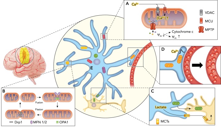
Schematic illustrating the response of astrocytic mitochondria to ischemia A: loss of oxygen and glucose leads to increased cytosolic Ca2+ concentrations that drives excessive accumulation of Ca2+ into the mitochondria via voltage-dependent anion channels (VDACs) and mitochondrial calcium uniporters (MCUs), triggering opening of the large mitochondrial permeability transition pore (MPTP). This allows the indiscriminate passage of small solutes out of the mitochondria causing dissipation of the mitochondrial membrane potential (ψm), which can culminate in release of cytochrome c and cellular apoptosis or membrane potential recovery with cell survival. B: mitochondria undergo morphological and network architectural changes in response to ischemia. They can adopt rounder discrete shape via fission mediated by dynamin-related protein 1 (Drp1) and fission protein 1 (Fis1), or form elongated, tubular interconnected network via fusion mediated by the outer membrane GTPases mitofusin-1 and -2 (MFN1/MFN2) and the inner membrane protein optic atrophy 1 (OPA1). C: astrocytes can promote neuronal survival by providing lactate as an energy substrate via monocarboxylate transporters (MCT) to neurons in the setting of impaired oxidative phosphorylation, in addition to directly donating functional mitochondria via a CD38-dependent mechanism. D: astrocyte mitochondria are enriched within vascular endfeet and may play a central role in neurovascular coupling by regulating Ca2+ signals. E: mitochondria are heterogeneous in structure and function, which may contribute to astroglial diversity. A subpopulation of astrocytic mitochondria may also be selectively resilient to ischemia.
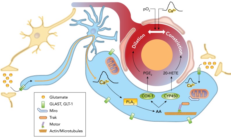
Schematic illustrating astrocytes as central mediators of neurovascular coupling Neuronal activity causes release of glutamate, which is taken up by astrocytes, triggering a Ca2+ signal that may be propagated down to or separately occurs within the vascular endfeet. This endfoot Ca2+ signal stimulates release of vasoactive factors, namely arachidonic acid (AA) and its metabolites—prostaglandins (PGs), epoxyeicosatrienoic acids (EETs), 20-hydroxyeicosatetraenoic acid (20-HETE)—onto cerebral blood vessels, evoking dilatation or constriction. The direction of blood vessel caliber change may be modulated by the partial pressure of oxygen in the blood (Po 2) or magnitude of the Ca2+ signal. The uptake of glutamate by astrocytes causes the immobilization of mitochondria near glutamate transporters in the processes or endfeet via changes in the binding of the transport proteins Miro and Trak. Astrocytic mitochondria are important in the generation and shaping of Ca2+ signals and thus likely play a key role in the control of blood flow in response to neuronal activity.
Similar articles
-
Astrocyte mitochondrial mechanisms of ischemic brain injury and neuroprotection.Neurochem Res. 2004 Mar;29(3):601-8. doi: 10.1023/b:nere.0000014830.06376.e6. Neurochem Res. 2004. PMID: 15038607 Review.
-
Astrocyte mitochondria in in vitro models of ischemia.J Bioenerg Biomembr. 2004 Aug;36(4):317-21. doi: 10.1023/B:JOBB.0000041761.61554.44. J Bioenerg Biomembr. 2004. PMID: 15377865 Review.
-
Brain-targeted heptapeptide-loaded exosomes attenuated ischemia-reperfusion injury by promoting the transfer of healthy mitochondria from astrocytes to neurons.J Nanobiotechnology. 2022 May 23;20(1):242. doi: 10.1186/s12951-022-01425-6. J Nanobiotechnology. 2022. PMID: 35606779 Free PMC article.
-
The Role of Astrocytic Mitochondria in the Pathogenesis of Brain Ischemia.Mol Neurobiol. 2024 Apr;61(4):2270-2282. doi: 10.1007/s12035-023-03714-z. Epub 2023 Oct 23. Mol Neurobiol. 2024. PMID: 37870679 Review.
-
No improvement of neuronal metabolism in the reperfusion phase with melatonin treatment after hypoxic-ischemic brain injury in the neonatal rat.J Neurochem. 2016 Jan;136(2):339-50. doi: 10.1111/jnc.13420. Epub 2015 Nov 24. J Neurochem. 2016. PMID: 26526584
Cited by
-
The Imbalance of Astrocytic Mitochondrial Dynamics Following Blast-Induced Traumatic Brain Injury.Biomedicines. 2023 Jan 24;11(2):329. doi: 10.3390/biomedicines11020329. Biomedicines. 2023. PMID: 36830865 Free PMC article.
-
Mitophagy-associated programmed neuronal death and neuroinflammation.Front Immunol. 2024 Oct 2;15:1460286. doi: 10.3389/fimmu.2024.1460286. eCollection 2024. Front Immunol. 2024. PMID: 39416788 Free PMC article. Review.
-
Nitroxidative Stress, Cell-Signaling Pathways, and Manganese Porphyrins: Therapeutic Potential in Neuropathic Pain.Int J Mol Sci. 2025 Feb 26;26(5):2050. doi: 10.3390/ijms26052050. Int J Mol Sci. 2025. PMID: 40076672 Free PMC article. Review.
-
Crosstalk between Neuron and Glial Cells in Oxidative Injury and Neuroprotection.Int J Mol Sci. 2021 Dec 10;22(24):13315. doi: 10.3390/ijms222413315. Int J Mol Sci. 2021. PMID: 34948108 Free PMC article. Review.
-
Differential Mitochondrial Bioenergetics in Neurons and Astrocytes Following Ischemia-Reperfusion Injury and Hypothermia.Biomedicines. 2024 Aug 1;12(8):1705. doi: 10.3390/biomedicines12081705. Biomedicines. 2024. PMID: 39200170 Free PMC article.
References
-
- Agarwal A, Wu PH, Hughes EG, Fukaya M, Tischfield MA, Langseth AJ, Wirtz D, Bergles DE. Transient opening of the mitochondrial permeability transition pore induces microdomain calcium transients in astrocyte processes. Neuron 93: 587–605.e7, 2017. doi:10.1016/j.neuron.2016.12.034. - DOI - PMC - PubMed
Publication types
MeSH terms
Grants and funding
LinkOut - more resources
Full Text Sources
Other Literature Sources