Review
. 2018 Mar;38(1):67-90.
doi: 10.1016/j.cll.2017.10.006.

Pathology-Based Research in Africa

Affiliations
Review

Pathology-Based Research in Africa

Maria P Lemos et al. Clin Lab Med. 2018 Mar.

Abstract

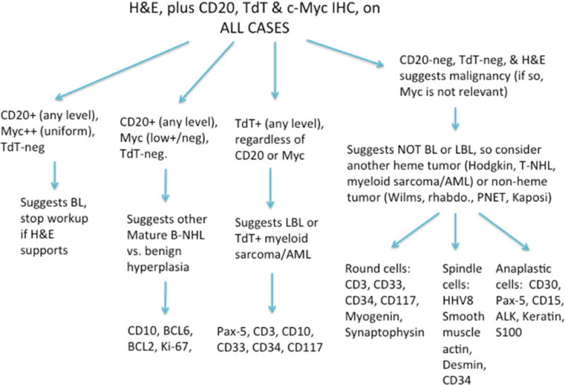
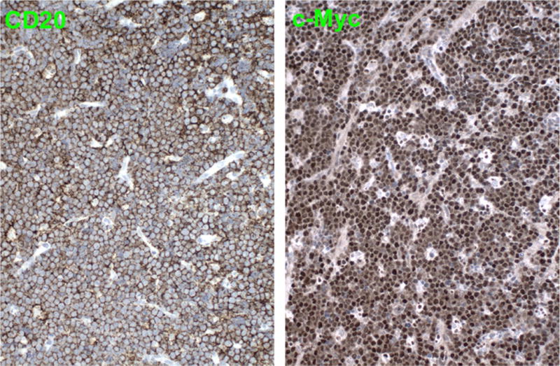
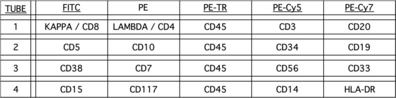
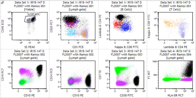
The process of conducting pathology research in Africa can be challenging. But the rewards in terms of knowledge gained, quality of collaborations, and impact on communities affected by infectious disease and cancer are great. This report reviews 3 different research efforts: fatal malaria in Malawi, mucosal immunity to HIV in South Africa, and cancer research in Uganda. What unifies them is the use of pathology-based approaches to answer vital questions, such as physiology, pathogenesis, predictors of clinical course, and diagnostic testing schemes.

Keywords: Burkitt lymphoma; Cancer; HIV/AIDS; Histopathology; Infectious disease; Malaria; Pathology.


Figures

FIGURE 1
FIGURE 1
Sampling proforma for autopsies performed for the study of clinico-pathological correlates of fatal cerebral malaria. Organs are listed by row. The first seven columns on the left represent samples destined for formalin fixation and paraffin embedding. Two samples (UBC vials and MAC) were preserved for electron microscopy. Tissue smears and touch preps were each prepared using 2 different approaches (air fixation, acetone fixation). Frozen samples (9 right most columns) were processed for expression analyses (in RNA later), IHC (in OCT), or were flash frozen.
FIGURE 2
FIGURE 2
A body prior to autopsy, wrapped in the traditional shroud (a local chitenge), on the plinth of the mortuary at the Queen Elizabeth Central Hospital in Blantyre, Malawi.
FIGURE 3
FIGURE 3
Within 2h of culture, injected IgG accumulates at the basal membrane of the epidermis in foreskin explants. Within 6h of collection, foreskin tissue explants from 3 men undergoing VMMC were sectioned, injected with antibodies (human IgG-Ax647), and cultured for 2h to observe the areas of antibody diffusion. Control samples received no antibody in their injection, or antibodies conjugated to another fluorochrome. Samples were fixed in formalin for 24h, and rinsed in water three times, rehydrated in two baths of 10% sucrose in 1X PBS, followed by three baths of 1X PBS, and embedded in OCT. After sectioning of the slides, samples were stained with anti-CD138 stain and biotin-conjugated goat anti-mouse IgG1 F(ab’)2 and avidin-AlexaFluor568 (pseudocolored red). All slides were counterstained with DAPI.
FIGURE 4
FIGURE 4
Within 24h of foreskin explant culture, injected IgG can be identified in epithelial cells. Within 6h of collection, foreskin tissue explants from 3 men undergoing VMMC was sectioned, injected with antibodies (human IgG-Ax647), and cultured for 24h to observe the areas of antibody diffusion. Control samples received no antibody in their injection, or antibodies conjugated to another fluorochrome. Samples were stained as indicated in Figure 3. CD138 was lost from the explant’s epithelium at long incubation times (24h), suggesting some ex-vivo modifications. IgG deposited at epithelial basal membrane and dermis, with sparse cells.
Figure 5
Figure 5
Figure 5 A demonstrates the IHC algorithm tested on pediatric small blue round cell tumors from SSA. Because BL is the most common pediatric malignancy in SSA, the initial round of IHC was targeted to BL, and included antibodies to CD20, TdT, and c-MYC. When these three stains did not confirm BL, several second-tier IHC combinations were performed, according to the details of the algorithm. The application of this algorithm to roughly 65 pediatric SBRCT cases suggested that TdT IHC is expendable for reliable diagnosis of BL, allowing this diagnosis to be made with near certainty with only 3 slides: an H&E, and CD20 and c-MYC IHC. The lower 2 figures (Figure 5B) show low power (40×) and high power (400×) views of a representative, very small, 2 to 3 mm in oral biopsy from a pediatric BL patient in the patient series. The 40× view shows an atypical diffuse lymphoid infiltrate, which at 400× has the characteristic appearance of BL, including medium-sized tumor cells and a “starry sky” appearance. Importantly, using only CD20 and c-MYC IHC (Figure 5C), the diagnosis of BL can be rendered with nearly 100% sensitivity and specificity: the uniform CD20 supports a mature B cell process, while the uniform strong c-Myc protein expression strongly suggests the c-MYC gene-containing chromosome 8 translocation characteristic of eBL. Additional studies on this series confirmed the expected presence of EBV infection (positive EBER-1 in situ hybridization) and the expected absence of lymphoblastic differentiation (negative TdT IHC).
Figure 5
Figure 5
Figure 5 A demonstrates the IHC algorithm tested on pediatric small blue round cell tumors from SSA. Because BL is the most common pediatric malignancy in SSA, the initial round of IHC was targeted to BL, and included antibodies to CD20, TdT, and c-MYC. When these three stains did not confirm BL, several second-tier IHC combinations were performed, according to the details of the algorithm. The application of this algorithm to roughly 65 pediatric SBRCT cases suggested that TdT IHC is expendable for reliable diagnosis of BL, allowing this diagnosis to be made with near certainty with only 3 slides: an H&E, and CD20 and c-MYC IHC. The lower 2 figures (Figure 5B) show low power (40×) and high power (400×) views of a representative, very small, 2 to 3 mm in oral biopsy from a pediatric BL patient in the patient series. The 40× view shows an atypical diffuse lymphoid infiltrate, which at 400× has the characteristic appearance of BL, including medium-sized tumor cells and a “starry sky” appearance. Importantly, using only CD20 and c-MYC IHC (Figure 5C), the diagnosis of BL can be rendered with nearly 100% sensitivity and specificity: the uniform CD20 supports a mature B cell process, while the uniform strong c-Myc protein expression strongly suggests the c-MYC gene-containing chromosome 8 translocation characteristic of eBL. Additional studies on this series confirmed the expected presence of EBV infection (positive EBER-1 in situ hybridization) and the expected absence of lymphoblastic differentiation (negative TdT IHC).
Figure 6
Figure 6
Four-tube, five-color flow cytometric assay designed to detect all hematopoietic neoplasms. In this assay, a PE-Texas Red-conjugated CD45 antibody is included in each tube, to ensure comparability of cell identification (gating) across the tubes when used in conjunction with SSC and FSC. Tube 1 takes advantage of the mutually exclusive nature of immunoglobulin light chain (kappa/lambda) expression on B cells and CD4/CD8 expression on T cells, which allows this tube to contain FITC-conjugated kappa and CD8 antibodies, and PE-conjugated lambda and CD4 antibodies in the same tube. The CD3 and CD20 antibodies in this tube allow initial separation of T and B cells, respectively, among which kappa:lambda and CD4:CD8 ratios can be respectively determined. Figure 6B shows the proof of concept for BL diagnosis by the four-tube, five-color flow assay. The flow histograms (dot plots) in this figure show an experiment in which the BL cell line Ramos was added to benign pleural fluid at roughly 25% of the total cells. The flow assay easily identifies the tumor cells, which are colored black throughout the figure. As expected for BL, the tumor cells expressed the leukocyte-common antigen CD45, the pan-B cell antigens CD19, CD20, and HLA-DR, and the germinal center B cell antigens CD10 and CD38; the tumor cells lambda light chain-restriction, confirming monoclonality, and increased forward scatter (FS INT) indicating increased cell size.
Figure 6
Figure 6
Four-tube, five-color flow cytometric assay designed to detect all hematopoietic neoplasms. In this assay, a PE-Texas Red-conjugated CD45 antibody is included in each tube, to ensure comparability of cell identification (gating) across the tubes when used in conjunction with SSC and FSC. Tube 1 takes advantage of the mutually exclusive nature of immunoglobulin light chain (kappa/lambda) expression on B cells and CD4/CD8 expression on T cells, which allows this tube to contain FITC-conjugated kappa and CD8 antibodies, and PE-conjugated lambda and CD4 antibodies in the same tube. The CD3 and CD20 antibodies in this tube allow initial separation of T and B cells, respectively, among which kappa:lambda and CD4:CD8 ratios can be respectively determined. Figure 6B shows the proof of concept for BL diagnosis by the four-tube, five-color flow assay. The flow histograms (dot plots) in this figure show an experiment in which the BL cell line Ramos was added to benign pleural fluid at roughly 25% of the total cells. The flow assay easily identifies the tumor cells, which are colored black throughout the figure. As expected for BL, the tumor cells expressed the leukocyte-common antigen CD45, the pan-B cell antigens CD19, CD20, and HLA-DR, and the germinal center B cell antigens CD10 and CD38; the tumor cells lambda light chain-restriction, confirming monoclonality, and increased forward scatter (FS INT) indicating increased cell size.

References

    1. UNAIDS. The Gap Report. UNAIDS; Geneva: 2014.
    1. UNAIDS. Global AIDS update 2016. UNAIDS; Geneva: 2016.
    1. World Malaria report 2015. World Health Organization; 2015.
    1. Ferlay J, Shin HR, Bray F, Forman D, Mathers C, Parkin DM. Estimates of worldwide burden of cancer in 2008: GLOBOCAN 2008. Int J Cancer. 2010;127(12):2893–2917. - PubMed
    1. Mathers CD, Loncar D. Projections of global mortality and burden of disease from 2002 to 2030. PLoS Med. 2006;3(11):e442. - PMC - PubMed