Review
. 2018 Apr:49:85-93.
doi: 10.1016/j.sbi.2018.01.008. Epub 2018 Feb 4.

Assembly and structure of the SSU processome-a nucleolar precursor of the small ribosomal subunit

Affiliations
Review

Assembly and structure of the SSU processome-a nucleolar precursor of the small ribosomal subunit

Jonas Barandun et al. Curr Opin Struct Biol. 2018 Apr.

Abstract

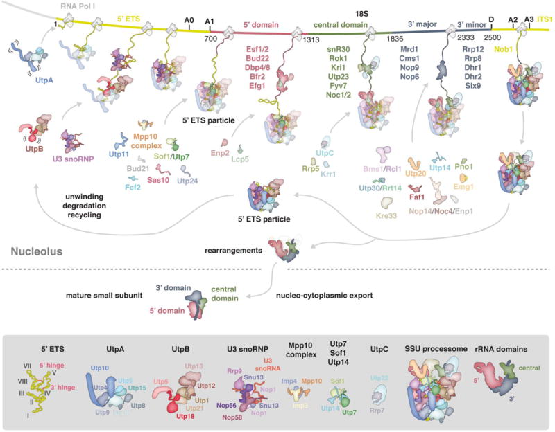
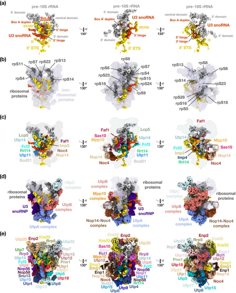
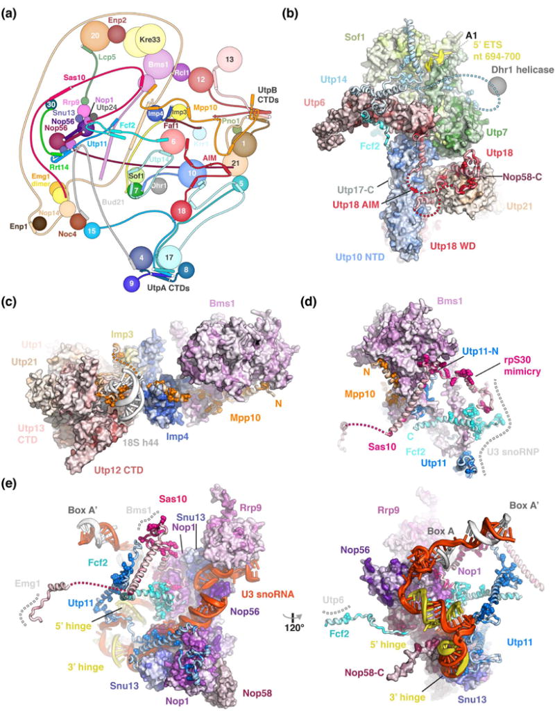
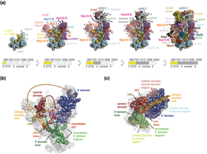
The small subunit processome is the first precursor of the small eukaryotic ribosomal subunit. During its assembly in the nucleolus, many ribosome biogenesis factors, an RNA chaperone, and ribosomal proteins associate with the nascent pre-rRNA. Biochemical studies have elucidated the rRNA-subdomain dependent recruitment of these factors during SSU processome assembly and have been complemented by structural studies of the assembled particle. Ribosome biogenesis factors encapsulate and guide subdomains of pre-ribosomal RNA in distinct compartments. This prevents uncoordinated maturation and enables processing of regions not accessible in the mature subunit. By sequentially reducing conformational freedom, flexible proteins facilitate the incorporation of dynamic subcomplexes into a globular particle. Large rearrangements within the SSU processome are required for compaction into the mature small ribosomal subunit.


Conflict of interest statement

Conflict of Interest

Nothing declared.

Figures

Figure 1
Figure 1
Co-transcriptional assembly of the small subunit processome. Schematic representation of early events of small subunit assembly in the nucleolus and subsequent maturation resulting in the mature cytoplasmic SSU. A section of the rDNA locus is shown with 5′ ETS and ITS1 colored in yellow and 18S colored in red (5′ domain), green (central domain) and slate (3′ domain). Factors visualized in the assembled SSU processome [30] are shown as schematic outline while transient components are listed and colored according to the rRNA domain they are associated with [19,20]. The SSU processome and its major components are shown in detail below.
Figure 2
Figure 2
Structural organization of the yeast small subunit processome. (a) RNA molecules of the SSU processome are shown as surfaces with 5′ ETS (yellow), U3 snoRNA (red) and pre-18S (light-grey). Structural elements of RNAs and helices of the 5′ ETS are indicated. (b) Ribosomal proteins are represented in dark-grey, non-ribosomal assembly factors in transparent light-blue, and RNA species as in (a). (c) Surface representation of centrally located ribosome assembly factors. (d) Visualization of the complexes UtpA (blue), UtpB (red), U3 snoRNP (purple), UtpC (light-blue), the Nop14-Noc4 complex (brown) and the Mpp10 complex (orange). (e) Surface representation of all individual components of the small subunit processome.
Figure 3
Figure 3
Peptides connect distant sites within the SSU processome via conserved binding motifs. (a) Schematic protein-protein interaction diagram of selected SSU processome components represented as spheres or lines with interacting elements as helices or strands. The Utps (U-three proteins) are labeled with their respective number. (b-e) Detailed views of Utp18 and Utp14 (b), Mpp10 (c), Bms1 (d) and the U3 snoRNP (e) with proteins shown as surface or cartoon, colored according to conservation with residues conserved more than 90 % highlighted as spheres. Direct interaction partners depicted as surface or a grey dashed line. All proteins are colored by conservation from lighter to darker shades. Clustal [49] was used to align manually curated sequences (H. sapiens, S. cerevisiae, G. gallus, D. melanogaster, S. pombe, C. elegans, D. rerio, A. thaliana, A. gambiae, P. troglodytes, R. norvegicus, M. musculus, B. taurus, S. scrofa) and plotted onto the structure using Homolmapper [50].
Figure 4
Figure 4
Three-dimensional maturation model of the Saccharomyces cerevisiae small-subunit processome. (a) Co-transcriptional assembly of the SSU processome as a function of transcription of rRNA regions (280 nucleotides of 5′ ETS, 5′ ETS, 5′ domain, central domain and 3′ domain). 5′ ETS (yellow), rRNA domains (white) and complexes such as UtpA (light-blue), UtpB (red), U3 snoRNP (purple, red), the Mpp10 complex (orange) and additional proteins, are shown as surfaces. Bound peptides are shown as cartoon with their initially flexible tails as dashed lines. As maturation progresses, these tails recruit additional factors and become ordered. The pre-rRNA shown in the intermediates is schematically indicated below each particle and colored in darker shades. As this model is based on the mature SSU processome structure, regions that are presumed to be flexible in earlier states are highlighted accordingly. (b, c) Comparative locations of rRNA domains within the SSU processome [PDB 5wlc] (b) and the mature small ribosomal subunit [pdb 4v88] (c). Individual rRNA domains are colored identically with 5′ domain (blue), central domain (red), 3′ domain (green) and shown as spheres superimposed onto transparent outlines of the particles. In the SSU processome, the flexible helix 44 is indicated as schematic outline. Rearrangements of rRNA domains from the SSU processome (b) that are necessary to obtain the positions within the mature small ribosomal subunit (c) are indicated with arrows. The central U3 snoRNA Box A and Box A′ are colored in purple. RNA elements disordered in the SSU processome are indicated in lighter shades in the mature SSU.

References

    1. Woolford JL, Baserga SJ. Ribosome Biogenesis in the Yeast Saccharomyces cerevisiae. Genetics. 2013;195:643–681. - PMC - PubMed
    1. Turowski TW, Tollervey D. Cotranscriptional events in eukaryotic ribosome synthesis. WIREs RNA. 2015;6:129–139. - PubMed
    1. Miller OL, Beatty BR. Visualization of nucleolar genes. Science. 1969;164:955–957. - PubMed
    1. Dragon F, Gallagher JEG, Compagnone-Post PA, Mitchell BM, Porwancher KA, Wehner KA, Wormsley S, Settlage RE, Shabanowitz J, Osheim Y, et al. A large nucleolar U3 ribonucleoprotein required for 18S ribosomal RNA biogenesis. Nature. 2002;417:967–970. - PMC - PubMed
    1. Grandi P, Rybin V, Bassler J, Petfalski E, Strauss D, Marzioch M, Schäfer T, Kuster B, Tschochner H, Tollervey D, et al. 90S pre-ribosomes include the 35S pre-rRNA, the U3 snoRNP, and 40S subunit processing factors but predominantly lack 60S synthesis factors. Mol Cell. 2002;10:105–115. - PubMed

Publication types

LinkOut - more resources