Review
. 2018 Feb 6;9(2):74.
doi: 10.3390/genes9020074.

Covalent Strategies for Targeting Messenger and Non-Coding RNAs: An Updated Review on siRNA, miRNA and antimiR Conjugates

Affiliations
Review

Covalent Strategies for Targeting Messenger and Non-Coding RNAs: An Updated Review on siRNA, miRNA and antimiR Conjugates

Santiago Grijalvo et al. Genes (Basel). .

Abstract

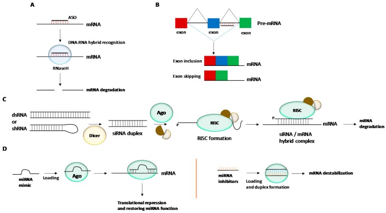
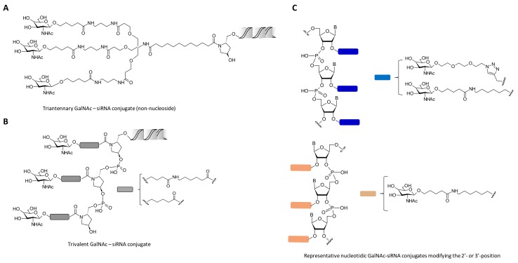
Oligonucleotide-based therapy has become an alternative to classical approaches in the search of novel therapeutics involving gene-related diseases. Several mechanisms have been described in which demonstrate the pivotal role of oligonucleotide for modulating gene expression. Antisense oligonucleotides (ASOs) and more recently siRNAs and miRNAs have made important contributions either in reducing aberrant protein levels by sequence-specific targeting messenger RNAs (mRNAs) or restoring the anomalous levels of non-coding RNAs (ncRNAs) that are involved in a good number of diseases including cancer. In addition to formulation approaches which have contributed to accelerate the presence of ASOs, siRNAs and miRNAs in clinical trials; the covalent linkage between non-viral vectors and nucleic acids has also added value and opened new perspectives to the development of promising nucleic acid-based therapeutics. This review article is mainly focused on the strategies carried out for covalently modifying siRNA and miRNA molecules. Examples involving cell-penetrating peptides (CPPs), carbohydrates, polymers, lipids and aptamers are discussed for the synthesis of siRNA conjugates whereas in the case of miRNA-based drugs, this review article makes special emphasis in using antagomiRs, locked nucleic acids (LNAs), peptide nucleic acids (PNAs) as well as nanoparticles. The biomedical applications of siRNA and miRNA conjugates are also discussed.

Keywords: RNA interference; covalent approach; gene delivery; gene therapy; miRNA technology; non-coding RNA; nucleic acid conjugates.


Conflict of interest statement

The authors declare no conflict of interest.

Figures

Figure 1
Figure 1
Gene regulation mechanisms mediated by antisense oligonucleotides (ASOs), triplex-forming oligonucleotides (TFOs), small interference RNAs (siRNAs) and microRNAs (miRNAs). (A) Antisense technology: RNAseH is able to recognize DNA:RNA hybrid complexes which leads to mRNA degradation and the blockade of protein synthesis. (B) RNA splicing mechanisms in order to restore the disrupted reading fragment of a gene by using ASOs targeting intron and exon junctions. (C) RNA interference: The siRNA duplex is unwound and the guide strand is recognized by Ago2. The resultant RISC complex is able to interact with complementary sequence of a target mRNA. (D) MiRNA technology: Gene expression can be modulated by using double or single-stranded miRNA mimics in order to restore the miRNA function by inhibiting translation process. MiRNA inhibitors with complementary sequences to miRNAs can also be used for inactivating ncRNAs.
Figure 2
Figure 2
Schemes of carbohydrate_siRNA conjugates containing one triantennary GalNAc (A), three consecutive GalNAc residues (B) and nucleotides carrying monovalent GalNAc units at different positions of the siRNA duplex (C). Modifications were introduced at the 3′-termini of the siRNA passenger strand.
Figure 3
Figure 3
(A) Chemical structure of polymer-siRNA conjugate_1. (B and C) Polymer-siRNA conjugate behaviours under extracellular and intracellular environments. SiRNA conjugate containing a PEG disulphide group (conjugate_2) liberated a siRNA molecule under extracellular conditions (B) whereas the nosyl group (conjugate_1) was able to liberate efficiently a siRNA molecule under intracellular conditions (combination of GSH and GST). (D) Gene silencing studies in the presence of lipofectamine in which shows the efficiency of conjugate_1 (containing the reducible nosyl group) to reduce significantly the production of luciferase. Adapted with permission from ref. [89]. Copyright 2017, Royal Society of Chemistry.
Figure 4
Figure 4
(A) Synthetic strategy for the preparation of SQ-siRNA NPs. (B) MTT assay at several concentrations of SQ-siRNA NPs and incubation times. (C) Reduction of RET/PTC1 mRNA expression in vitro after incubating siRNA RET/PTC1 (60 nM) in the presence of lipofectamine. (D) Effect of SQ-siRNA NPs in reducing the tumour growth after 20 days of treatment. (E) Reduction of RET/PTC21 mRNA expression measured by qRT-PCR after incubation with SQ-siRNA NPs in vivo. The silence activity mediated by SQ-siRNA NPs was significant (*) when compared to untreated cells according to ANOVA analysis. A scrambled siRNA sequence (control siRNA) was used in order to determine the specificity of the gene silencing process. Adapted with permission from ref. [118]. Copyright 2011, American Chemical Society.
Figure 5
Figure 5
(A) Synthetic strategy for the preparation of L-serine-based siRNA conjugates (green) modified with docosahexaenoic acid (DHA) (blue) at the 3’-termini of the passenger strand. (B) Confocal images at various incubation times (1, 4, 24 and 72 h) when DHA-siRNA and Chol-siRNA conjugates were incubated in the presence of neurons. (C) Reduction of Mtt mRNA expression mediated by DHA-siRNA and Chol-siRNA conjugates. Adapted with permission from ref. [126]. Copyright 2016, Nature Publishing Group.
Figure 6
Figure 6
(A) Chemical structure of Chol-siRNA conjugate containing the hydrophobic residue at the 5’-termini of the siRNA passenger strand. (B) Dynamic light scattering (DLS) of EVs alone and in the presence of Chol-siRNA conjugate. (C) Dose-response at several concentrations of EVs:siRNA (100, 200, 300, 400 and 600 nM) targeting HuR mRNA expression. (D) Evaluation of silencing process during time (24, 48, 72 and 168 h). * represents significant values of Chol-siRNA conjugate when compared to negative controls (untreated cells). Adapted with permission from ref. [133]. Copyright 2017, The American Society of Gene and Cell Therapy.
Figure 7
Figure 7
(A) Design of aptamer-siRNA chimeras targeting SLUG and NRP1 genes. (B,C) Inhibition of gene expression mediated by aptamer-siRNA chimeras targeting SLUG (B) and NRP1 (C) mRNA expressions. * represents significant values of the siRNA nanostructure when compared to untreated cells. Adapted with permission from ref. [156]. Copyright 2014, Elsevier.
Figure 8
Figure 8
Use of iron NPs functionalized with LNA targeting miR-10b. Structures of three NPs containing SPDP, GMBS and PEG linkers.
Figure 9
Figure 9
(A) Design and a TEM image of the AuNP conjugates decorated with VCAM1 binding peptide and anti-miR-712. (B) Confocal images of iMAECs which show the ability of the AuNPs to impart cellular uptake as well as the release of the fluorescently labelled anti-miR-712 and duplex formation with the corresponding target. This effect was not observed in non-expressing VCAM1 endothelial cells. Adapted with permission from [229]. Copyright 2016, Wiley-VCH Verlag GmbH&Co. KGaA, Weinheim.

References

    1. Doak B.C., Over B., Giordanetto F., Kihlberg J. Oral druggable space beyond the rule of 5: Insights from drugs and clinical candidates. Chem. Biol. 2014;21:1115–1142. doi: 10.1016/j.chembiol.2014.08.013. - DOI - PubMed
    1. Bennett C.F., Swayze E.E. RNA Targeting therapeutics: Molecular mechanisms of antisense oligonucleotides as a therapeutic platform. Annu. Rev. Pharmacol. Toxicol. 2010;50:259–293. doi: 10.1146/annurev.pharmtox.010909.105654. - DOI - PubMed
    1. Stephenson M.L., Zamecnik P.C. Inhibition of Rous sarcoma viral RNA translation by a specific oligodeoxyribonucleotide. Proc. Natl. Acad. Sci. USA. 1978;75:285–288. doi: 10.1073/pnas.75.1.285. - DOI - PMC - PubMed
    1. Matsui M., Corey D.R. Non-coding RNAs as drug targets. Nat. Rev. Drug Discov. 2016;16:167–179. doi: 10.1038/nrd.2016.117. - DOI - PMC - PubMed
    1. Hüttenhofer A., Schattner P., Polacek N. Non-coding RNAs: Hope or hype? Trends Genet. 2005;21:289–297. doi: 10.1016/j.tig.2005.03.007. - DOI - PubMed