. 2018 Feb 7;9(2):175.
doi: 10.1038/s41419-017-0204-3.

circHIPK3 regulates cell proliferation and migration by sponging miR-124 and regulating AQP3 expression in hepatocellular carcinoma

Affiliations

circHIPK3 regulates cell proliferation and migration by sponging miR-124 and regulating AQP3 expression in hepatocellular carcinoma

Genwen Chen et al. Cell Death Dis. .

Abstract

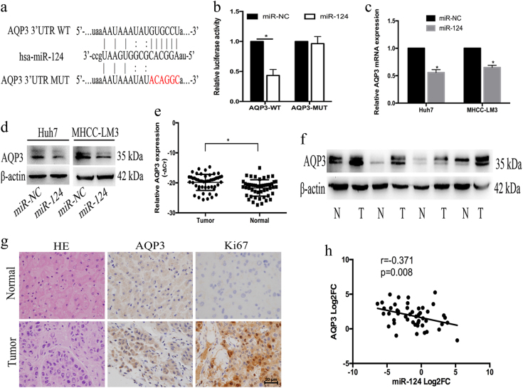
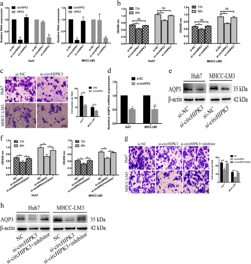
Noncoding RNAs plays an important role in hepatocellular carcinoma (HCC). Here, we show that miR-124 was downregulated in HCC tissues and that the ectopic expression of miR-124 inhibited the proliferation and migration of HCC cells. We proposed that aquaporin 3 (AQP3) is a direct target of miR-124. AQP3 was upregulated in HCC tissues and inversely correlated with miR-124 expression. The overexpression of miR-124 decreased AQP3 expression. Indeed, AQP3 overexpression promoted cell proliferation and migration, whereas miR-124 knockdown suppressed cell proliferation and migration. Furthermore, we found that circular RNA HIPK3 (circHIPK3) acted as a miR-124 sponge and regulated the expression of the miR-124 target gene AQP3. circHIPK3 was upregulated in HCC tissues and positively correlated with AQP3 expression. Thus, silencing circHIPK3 inhibited cell proliferation and migration by downregulating AQP3 expression. Moreover, miR-124 inhibition rescued circHIPK3 knockdown induced reduction in cell proliferation and migration, as well as AQP3 expression. In vivo experiments also confirmed that circHIPK3 regulated xenograft tumor growth via the miR-124-AQP3 axis. These observations indicate a possible novel therapeutic strategy involving circular RNAs in HCC.


Conflict of interest statement

The authors declare no competing financial interests.

Figures

Fig. 1
Fig. 1. miR-124 was downregulated in HCC and inhibited the proliferation and migration of HCC cells.
a Expression levels of miR-124 in HCC tissues and adjacent normal tissues (n = 50). b Expression levels of miR-124 in HCC and L02 cell lines. c CCK-8 assay after 72 h and 96 h treatment with miR-124 mimic in Huh7 and MHCC-LM3 cells. d The migration ability of Huh7 and MHCC-LM3 cells were measured by transwell assay (original magnification, ×200). Error bars represent mean ± SD from three independent experiments. *p < 0.05
Fig. 2
Fig. 2. AQP3 was a target of miR-124 and was upregulated in HCC.
a WT and MUT sequences designed for AQP3 according to the binding site for miR-124. b Relative luciferase activities after cotransfection pmirGLO-AQP3-WT or pmirGLO-AQP3-MUT with miR-124 mimic, respectively. c The mRNA expression of AQP3 was examined in Huh7 and MHCC-LM3 cells after transfection with miR-124 mimic. d The protein expression of AQP3 was examined by western blot in Huh7 and MHCC-LM3 cells after transfection with miR-124 mimic. e The AQP3 expression levels in HCC tissues and adjacent normal tissues by qRT-PCR (n = 50). f High expression of AQP3 was observed in HCC tissues by western blot. g Haematoxylin and eosin (H&E) and the expressions of AQP3 and Ki67 in HCC tissues and adjacent normal tissues were examined by IHC. h The inverse relationship between miR-124 and AQP3 in HCC tissues. Error bars represent mean ± SD from three independent experiments. *p < 0.05
Fig. 3
Fig. 3. AQP3 promoted the proliferation and migration of HCC cells.
a Knockdown efficacy of AQP3 in Huh7 and MHCC-LM3 cells by western blot analysis. b, c Knockdown of AQP3 suppressed the proliferation and migration in Huh7 and MHCC-LM3 cells determined by CCK-8 assay and transwell assay. d Overexpression efficacy of AQP3 in Huh7 and MHCC-LM3 cells by western blot. e, f AQP3 overexpression promoted the proliferation and migration in Huh7 and MHCC-LM3 cells by CCK-8 assay and transwell assay. Error bars represent mean ± SD from three independent experiments. *p < 0.05
Fig. 4
Fig. 4. Identification and characterization of circHIPK3 in HCC.
a Schematic representation of the target site in circHIPK3 for miR-124. b Schematic diagram of the head-to-tail splicing of circHIPK3 and Sanger sequencing of a PCR product. c Relative luciferase activities after cotransfection pmirGLO-circHIPK3-WT or pmirGLO-circHIPK3-MUT with miR-124 mimic, respectively. d, e The expression levels of circHIPK3 and HIPK3 mRNA in HCC tissues and adjacent normal tissues (n = 50). f The levels of HIPK3 mRNA were tightly correlated with circHIPK3. g The levels of AQP3 were positively correlated with circHIPK3. Error bars represent mean ± SD from three independent experiments. *p < 0.05
Fig. 5
Fig. 5. circHIPK3 regulated the proliferation and migration of HCC cells via the miR-124-AQP3 axis.
a Knockdown efficacy of circHIPK3 and HIPK3 mRNA in Huh7 and MHCC-LM3 cells was determined by qRT-PCR, respectively. b The effects of circHIPK3 and HIPK3 mRNA knockdown on Huh7 and MHCC-LM3 cells by CCK-8 assay. (c) circHIPK3 knockdown suppressed the migration of Huh7 and MHCC-LM3 cells. d, e circHIPK3 knockdown decreased AQP3 mRNA and protein expression in Huh7 and MHCC-LM3 cells. f, g miR-124 inhibitor rescued si-circHIPK3-induced reduction of the proliferation and migration in both Huh7 and MHCC-LM3 cells. h Reduction of AQP3 induced by si-circHIPK3 could be rescued by miR-124 inhibition. Error bars represent mean ± SD from three independent experiments. *p < 0.05; ns: no significance
Fig. 6
Fig. 6. Knockdown of circHIPK3 suppressed tumor growth in vivo.
a The xenograft tumors were significantly depressed by circHIPK3 depletion. b, c Tumor volume and tumor weight of the xenografts were remarkably inhibited by sh-circHIPK3. d circHIPK3 depletion downregulated AQP3 expression in vivo by western blot analysis. e The expressions of AQP3 and Ki67 in the xenograft tumors were examined by IHC. Error bars represent mean ± SD from three independent experiments. *p < 0.05

References

    1. Torre LA, et al. Global Cancer Statistics, 2012. CA Cancer J. Clin. 2015;65:87–108. doi: 10.3322/caac.21262. - DOI - PubMed
    1. Chen W, et al. Cancer Statistics in China, 2015. CA Cancer J. Clin. 2016;66:115–132. doi: 10.3322/caac.21338. - DOI - PubMed
    1. Agre P. The aquaporin water channels. Proc. Am. Thorac. Soc. 2006;3:5–13. doi: 10.1513/pats.200510-109JH. - DOI - PMC - PubMed
    1. Magni F, et al. Proteomic knowledge of human aquaporins. Proteomics. 2006;6:5637–5649. doi: 10.1002/pmic.200600212. - DOI - PubMed
    1. Hu J, Verkman AS. Increased migration and metastatic potential of tumor cells expressing aquaporin water channels. FASEB J. 2006;20:1892. doi: 10.1096/fj.06-5930fje. - DOI - PubMed

Publication types

MeSH terms