. 2017 Nov 23;9(1):539-552.
doi: 10.18632/oncotarget.22640. eCollection 2018 Jan 2.

Loss of epithelium-specific GPx2 results in aberrant cell fate decisions during intestinal differentiation

Affiliations

Loss of epithelium-specific GPx2 results in aberrant cell fate decisions during intestinal differentiation

Claudia Lennicke et al. Oncotarget. .

Abstract

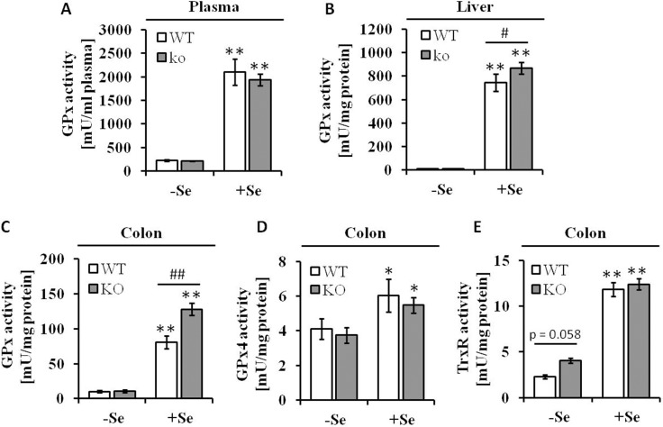
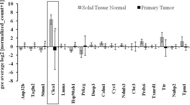
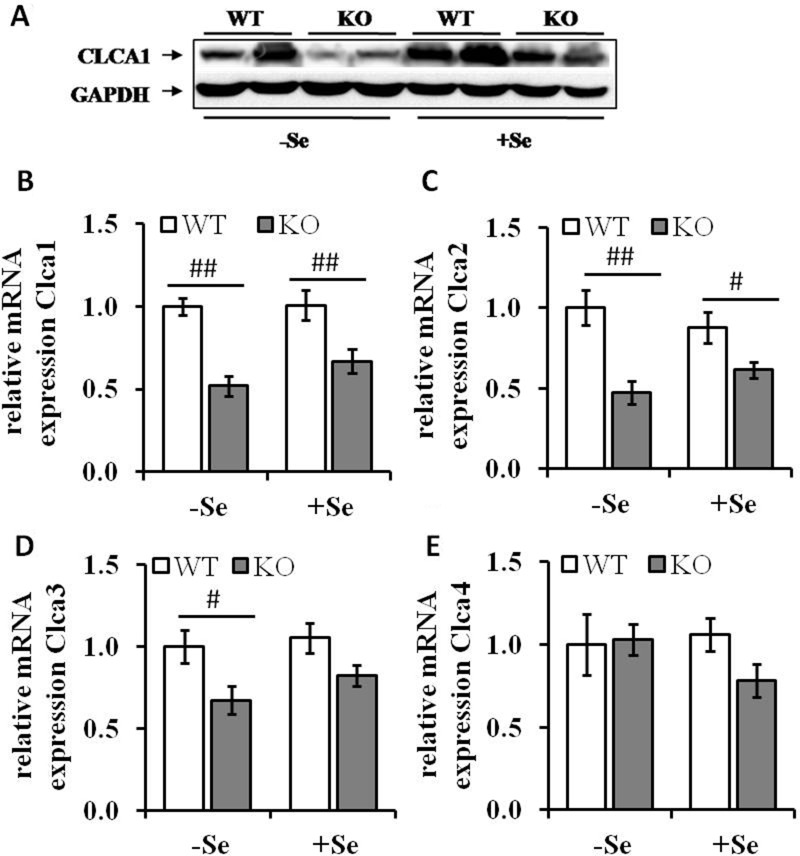
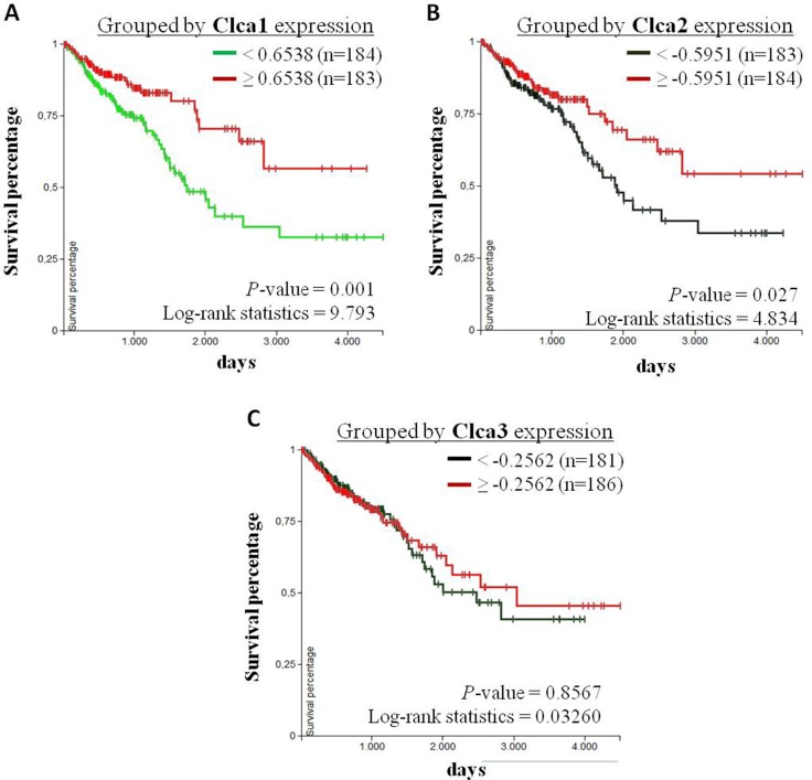
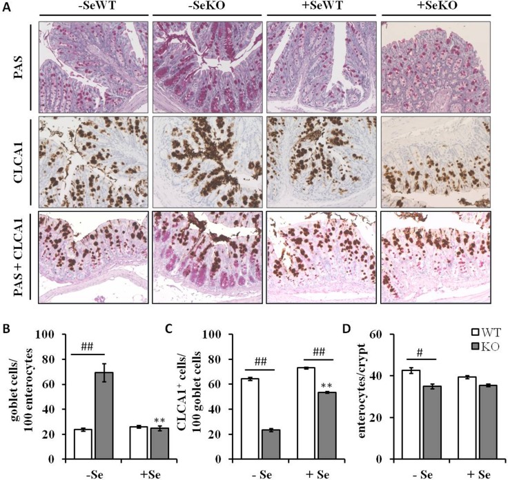
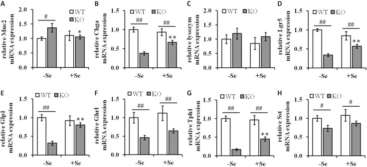
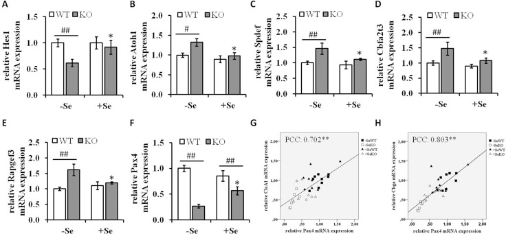
The selenoprotein glutathione peroxidase 2 (GPx2) is expressed in the epithelium of the gastrointestinal tract, where it is thought to be involved in maintaining mucosal homeostasis. To gain novel insights into the role of GPx2, proteomic profiles of colonic tissues either derived from wild type (WT) or GPx2 knockout (KO) mice, maintained under selenium (Se) deficiency or adequate Se supplementation conditions were established and analyzed. Amongst the panel of differentially expressed proteins, the calcium-activated chloride channel regulator 1 (CLCA1) was significantly down-regulated in GPx2 KO versus WT mice regardless of the given Se status. Moreover, transcript levels of the isoforms CLCA2 and CLCA3 showed a similar expression pattern. In the intestine, CLCA1 is usually restricted to mucin-producing goblet cells. However, although -SeKO mice had the highest numbers of goblet cells as confirmed by significantly enhanced mRNA expression levels of the goblet cell marker mucin-2, the observed expression pattern suggests that GPx2 KO goblet cells might be limited in synthesizing CLCA1. Furthermore, transcript levels of differentiation markers such as chromogranin-1 (Chga) for enteroendocrine cells and leucine-rich repeat-containing G-protein coupled receptor 5 (Lgr5) for stem cells were also downregulated in GPx2 KO mice. Moreover, this was accompanied by a downregulation of the mRNA expression levels of the intestinal hormones glucagon-like peptide 1 (Glp1), ghrelin (Ghrl) and somatostatin (Sst). Thus, it seems that GPx2 might be important for the modulation of cell fate decisions in the murine intestinal epithelium.

Keywords: Clca1; DIGE; glutathione peroxidase 2; selenium; stem cells.


Conflict of interest statement

CONFLICTS OF INTEREST The authors would like to state that they have no potential conflicts of interest to report.

Figures

Figure 1
Figure 1. Se status following eight weeks of feeding with different Se concentrations
Enzyme activities of (A) GPx in plasma, (B) GPx in liver and (C) in colon lysates, (D) GPx4 and (E) TrxR in colon tissues were spectrophotometrically determined as described in the Material and Methods section. Values are given as means ± S.E.M. (n = 9). Significant differences were calculated by one-way ANOVA. *p < 0.05; **p < 0.01 vs. the respective -Se group; ##p < 0.01 WT vs. KO within the same Se supply level.
Figure 2
Figure 2. Gene expression pattern of potential targets defined via the murine proteomic profiling experiments in CRC patients
Analysis of the expression pattern of proteins, which were found to be differentially regulated by selenium and/or the GPx2 status in the murine model system (see also Table 1) in regard to their potential role in CRC patients (TCGA-COADREAD; n = 434). Expression data were Log2 transformed, normalized TCGA-COADREAD data were grouped by the sample types and expressed as means ± S.D. (solid tissue normal n = 54, primary tumor n = 380). *p < 0.05, student´s t-test.
Figure 3
Figure 3. Expression levels of Clca isoforms are decreased by GPx2 knockout
(A) Protein levels of CLCA1 were analyzed by Western blotting; mRNA expression levels of (B) Clca1, (C) Clca2, (D) Clca3 and (E) Clca4 were analyzed by qRT-PCR, normalized to the amplification data of GAPDH, ß-actin and RPL13a and expressed in relation to the –SeWT group. Values are given as means ± S.E.M (n = 9 per group). #p < 0.05, ##p < 0.01 (one-way ANOVA).
Figure 4
Figure 4. Correlation analysis of Clca isoform expression levels with overall survival of CRC patients
Kaplan-Meier curves of CRC patients with primary tumors (TCGA-COADREAD, n = 380) were analyzed using the UCSC Xena browser (https://xenabrowser.net). Patients were grouped by the 50 percentiles of (A) Clca1, (B) Clca2 and (C) Clca3 expression levels. The differences between the curves were analyzed with the log-rank test and considered to be statistically significant when p < 0.05.
Figure 5
Figure 5. Histological and immunohistochemical analyses of colon sections
(A) Representative microscopy pictures of colon sections stained with PAS for counting goblet cells (upper row), an anti-CLCA1 antibody for visualizing CLCA1 positive cells (middle row), and co-staining of goblet cells and CLCA1 positive cells (bottom row) (B) number of goblet cells per 100 enterocytes, (C) number of CLCA1 positive cells per 100 goblet cells and (D) number of enterocytes per colon crypt are expressed as means ± S.E.M (n = 4 per group). *p < 0.05, **p < 0.01 vs. the respective -Se group; #p < 0.05, ##p < 0.01 WT vs. KO within the same Se supply (one-way ANOVA). HPF, high power field, CLCA1, Calcium-activated chloride channel regulator 1; PAS, periodic acid-Schiff.
Figure 6
Figure 6. Analysis of differentiation markers in colon lysates of GPx2 KO mice
mRNA expression levels of differentiation markers of (A) Muc2 (goblet cells), (B) Chga (enteroendocrine cells), (C) lysozyme (paneth like cells) and (D) Lgr5 (stem cells) and the mRNA expression pattern of the intestinal hormones, (E) Glp1, (F) Ghrl, (G) Tph1 and (H) Sst were determined by qPCR, normalized to the amplification data of GAPDH, ß-actin and RPL13a and expressed in relation to the -SeWT group. Data are given as means ± S.E.M. (n = 9 per group). *p < 0.05, **p < 0.01 vs. the respective -Se group; #p < 0.05, ##p < 0.01 WT vs KO within the same Se supply (one-way ANOVA). Muc2, mucin-2; Chga, chromogranin A; Lgr5, Leucine-rich repeat-containing G-protein coupled receptor 5. Glp1, glucagon-like peptide 1; Ghrl, ghrelin; Tph1, tryptophan 5-hydroxylase 1; Sst, somatostatin.
Figure 7
Figure 7. mRNA expression of components involved in differentiation processes
mRNA expression levels of the Notch signaling components (A) Hes1, (B) Atoh1, the Atoh1 downstream targets (C) Spdef, (D) Cbfa2t3, (E) Rapgef3 as well as of the transcription factor (F) Pax4 were determined by qRT-PCR normalized to the amplification data of GAPDH, ß-actin and RPL13a and expressed in relation to the –SeWT group. Data are expressed as means ± S.E.M. (n = 9 per group). *p < 0.05, **p < 0.01 vs. the respective -Se group; #p < 0.05, ##p < 0.01 WT vs KO within the same Se supply (one-way ANOVA). Correlation analysis of the expression pattern of Pax4, (G) Clca1 and (H) Chga were conducted by determining the Pearson Correlation Coefficients (PCC). **indicate significant correlations with a p-value < 0.01. Atoh1, protein atonal homolog 1; CLCA1, calcium-activated chloride channel regulator 1; Hes1, hairy and enhancer of split-1; Pax4, paired box protein Pax-4.

References

    1. Wessjohann LA, Schneider A, Abbas M, Brandt W. Selenium in chemistry and biochemistry in comparison to sulfur. Biol Chem. 2007;388:997–1006. https://doi.org/10.1515/BC.2007.138. - DOI - PubMed
    1. Steinbrenner H, Speckmann B, Klotz LO. Selenoproteins: Antioxidant selenoenzymes and beyond. Arch Biochem Biophys. 2016;595:113–9. https://doi.org/10.1016/j.abb.2015.06.024. - DOI - PubMed
    1. Varlamova EG, Cheremushkina IV. Contribution of mammalian selenocysteine¬containing proteins to carcinogenesis. Journal of Trace Elements in Medicine and Biology. 2017:76–85. https://doi.org/10.1016/j.jtemb.2016.08.004. - DOI - PubMed
    1. Duffield-Lillico AJ, Reid ME, Turnbull BW, Combs Jr GF, Slate EH, Fischbach LA, Marshall JR, Clark LC. Baseline characteristics and the effect of selenium supplementation on cancer incidence in a randomized clinical trial: a summary report of the Nutritional Prevention of Cancer Trial. Cancer Epidemiol Biomarkers &amp; Prev. 2002;11:630–9. - PubMed
    1. Micke O, Schomburg L, Buentzel J, Kisters K, Muecke R. Selenium in oncology: From chemistry to clinics. Molecules. 2009:3975–88. https://doi.org/10.3390/molecules14103975. - DOI - PMC - PubMed

LinkOut - more resources