

Folate receptor-α targeted near-infrared fluorescence imaging in high-risk endometrial cancer patients: a tissue microarray and clinical feasibility study
- PMID: 29416655
- PMCID: PMC5787511
- DOI: 10.18632/oncotarget.23155
Folate receptor-α targeted near-infrared fluorescence imaging in high-risk endometrial cancer patients: a tissue microarray and clinical feasibility study
Abstract
Objective: Detection and resection of all malignant lesions is pivotal in staging and cytoreductive surgery (CRS) of endometrial cancer (EC). Intraoperative EC detection could be enhanced using OTL-38, a fluorescent-labelled folate receptor-α (FRα) targeted imaging agent. The objectives of this study were to investigate which subgroups of high-risk EC patients express FRα and assess feasibility of intraoperative EC detection using OTL-38.
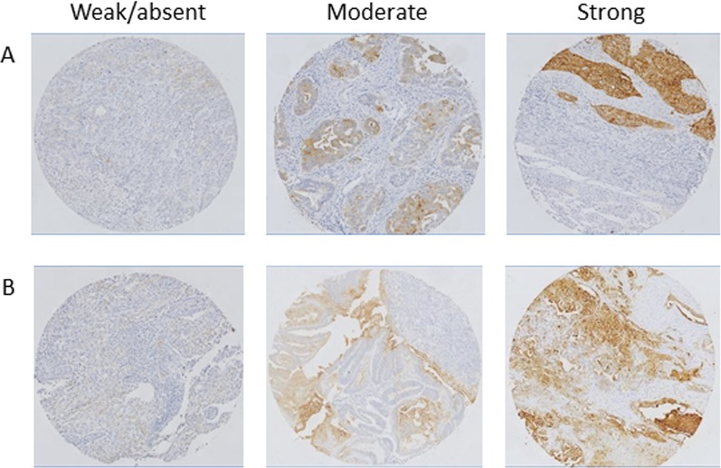
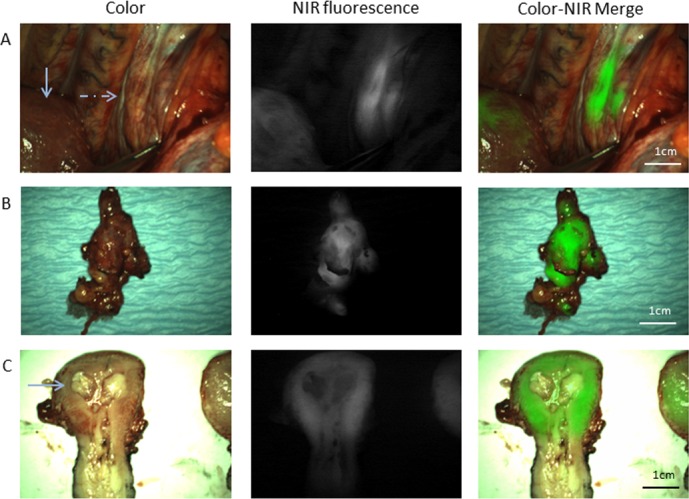
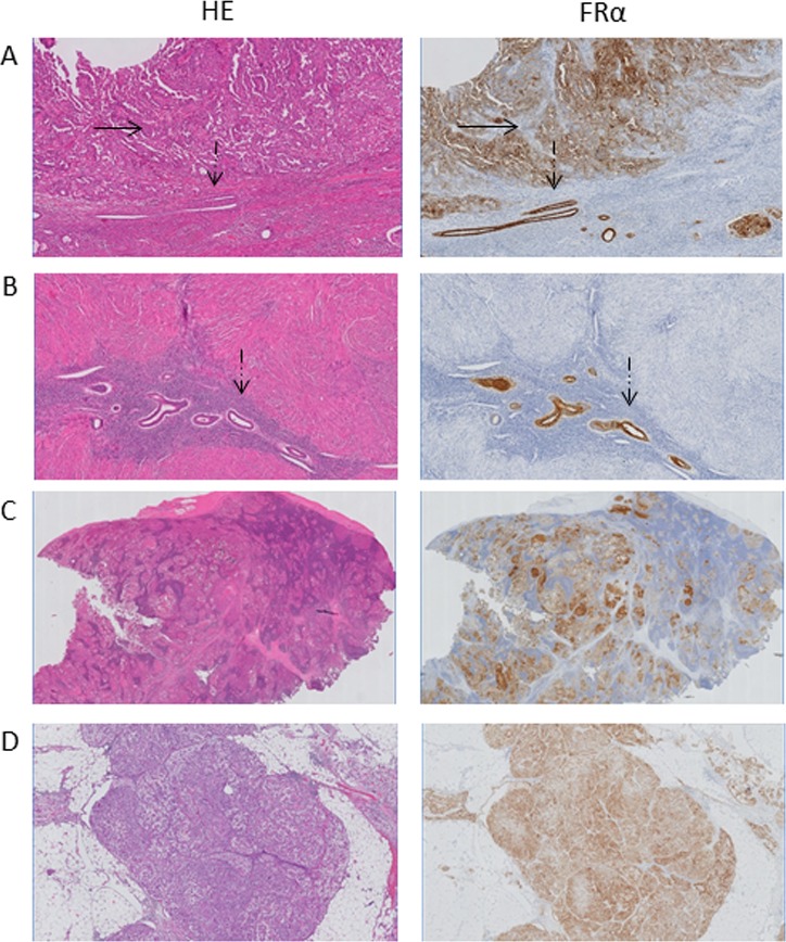
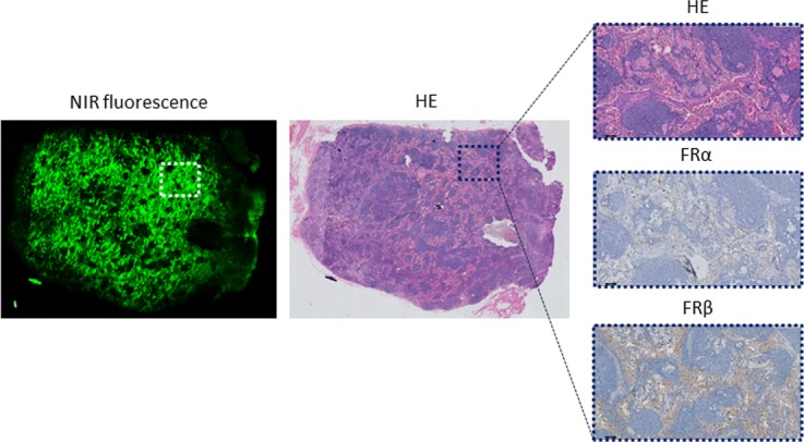
Results: FRα expression on TMA was significantly correlated with tumor type (p < 0.01). Eighty-two percent of serous and clear cell carcinomas showed FRα expression. Four patients were enrolled in the clinical study. Using fluorescence imaging all omental (n = 3) and lymph node (LN) metastases (n = 16) could be clearly identified, including one otherwise undetected omental metastasis. However, false-positive fluorescence was identified in 17/50 non-metastatic LNs, caused by OTL-38 targeting of FRβ, expressed by tumor-associated activated macrophages.
Conclusions: This study describes high FRα expression in serous and clear cell EC and demonstrates the first experience of intraoperative FRα-targeted tumor detection in patients with these subtypes of EC. Although all metastases could be clearly identified using OTL-38, the role of tumor-associated macrophages should be further evaluated.
Methods: Immunohistochemical (IHC) staining of FRα expression was performed on tissue micro arrays (TMA) of 116 patients with high-risk EC features. Patients with either serous or clear cell EC, planned for staging or CRS, were eligible for inclusion in the clinical study and received an intravenous dose of 0.0125 mg/kg OTL-38, 2-3 hours prior to surgery. Resected lesions, identified by standard-of-care and/or fluorescence imaging, were histopathologically assessed for FRα and tumor status.
Keywords: FRα; biomarker; targeting; tumor-specific imaging.
Conflict of interest statement
CONFLICTS OF INTEREST The Centre for Human Drug Research (a not-for-profit foundation) and the Leiden University Medical Center received financial compensation, study drug and equipment for the execution of this study from On Target Laboratories LLC.
Figures
References
-
- Amant F, Moerman P, Neven P, Timmerman D, Van Limbergen E, Vergote I. Endometrial cancer. Lancet. 2005;366:491–505. - PubMed
-
- Bansal N, Yendluri V, Wenham RM. The molecular biology of endometrial cancers and the implications for pathogenesis, classification, and targeted therapies. Cancer Contr. 2009;16:8–13. - PubMed
-
- Stelloo E, Bosse T, Nout RA, MacKay HJ, Church DN, Nijman HW, Leary A, Edmondson RJ, Powell ME, Crosbie EJ, Kitchener HC, Mileshkin L, Pollock PM, et al. Refining prognosis and identifying targetable pathways for high-risk endometrial cancer; a TransPORTEC initiative. Mod Pathol. 2015;28:836–844. - PubMed
LinkOut - more resources
Full Text Sources
Other Literature Sources
