. 2017 Dec 19;9(1):1210-1228.
doi: 10.18632/oncotarget.23456. eCollection 2018 Jan 2.

KSHV LANA upregulates the expression of epidermal growth factor like domain 7 to promote angiogenesis

Affiliations

KSHV LANA upregulates the expression of epidermal growth factor like domain 7 to promote angiogenesis

Suhani Thakker et al. Oncotarget. .

Abstract

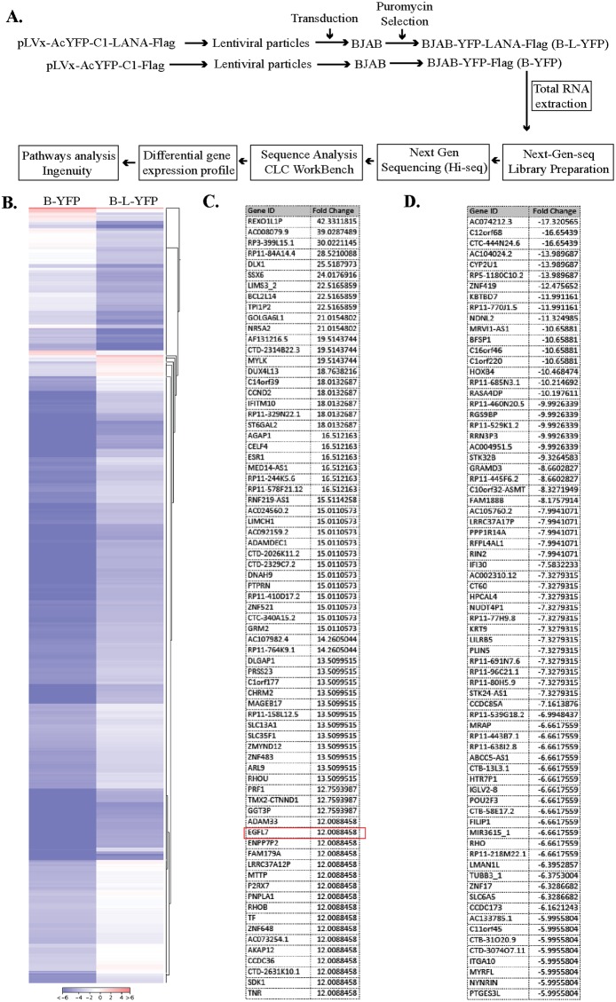
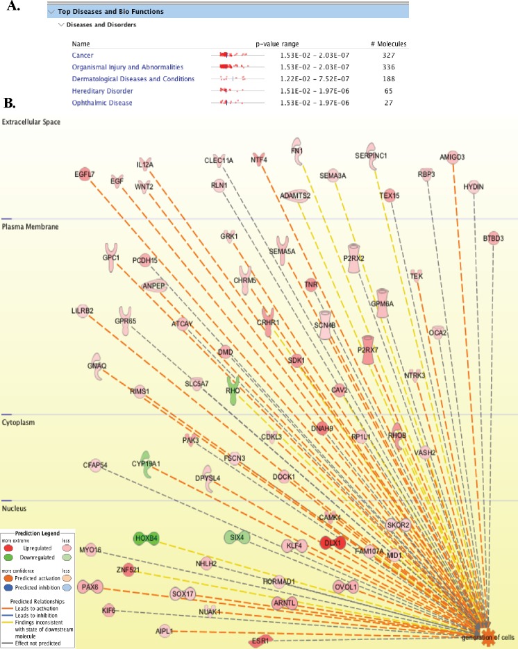
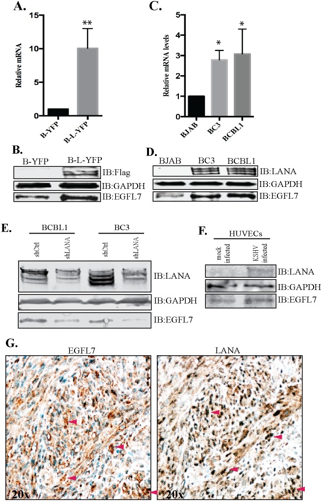
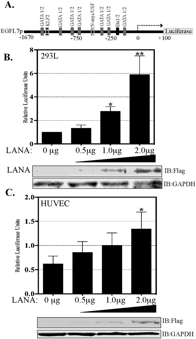
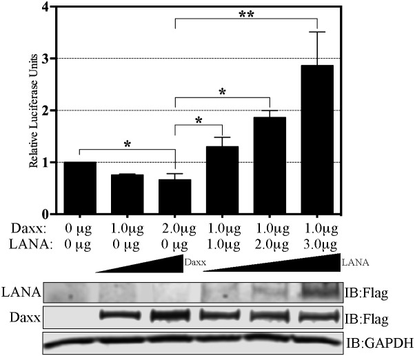
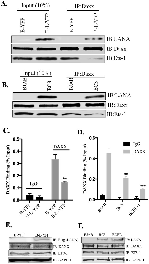
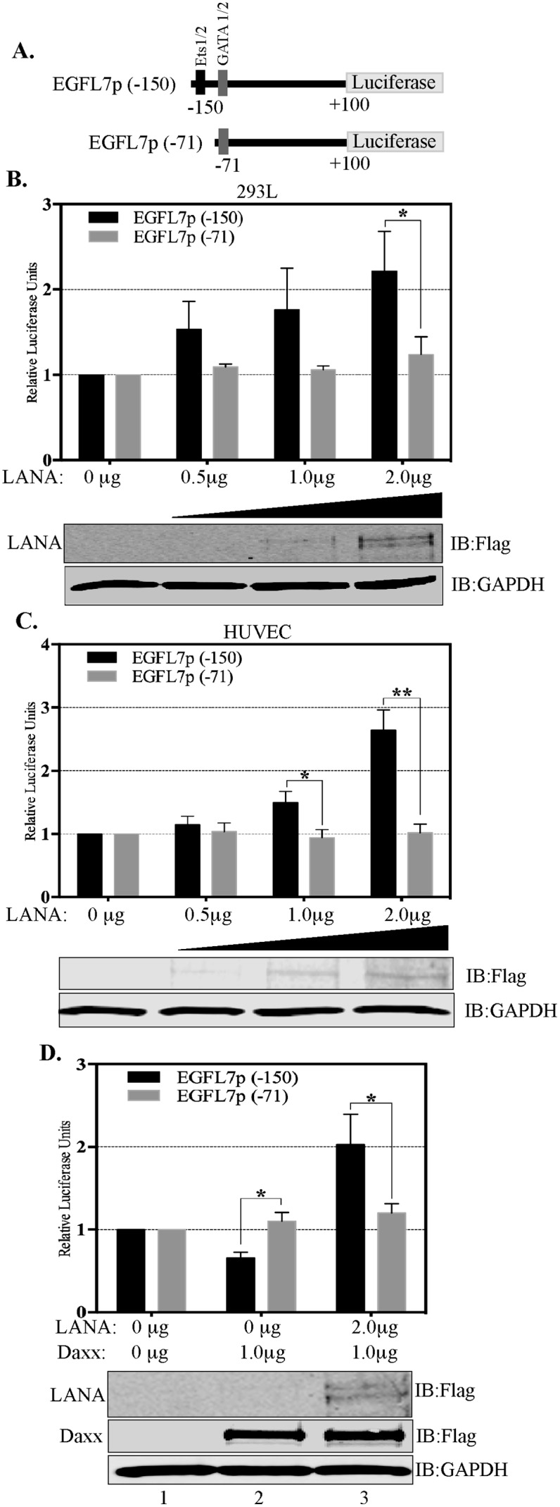
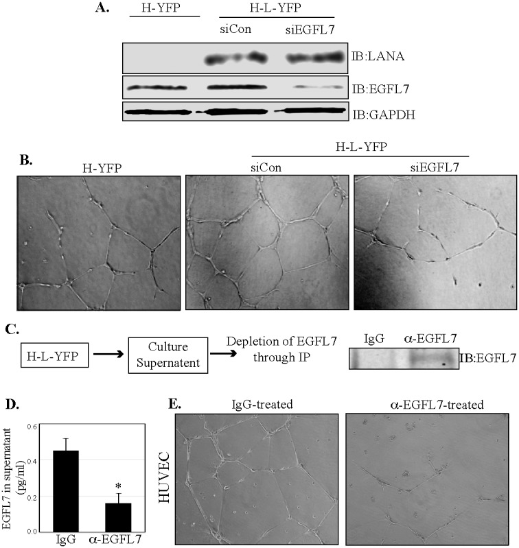
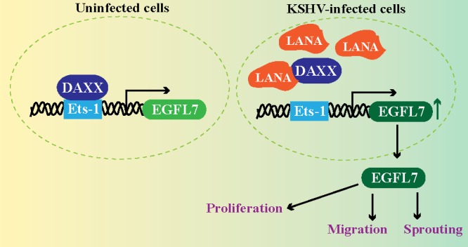
Kaposi's sarcoma (KS) is a highly-vascularized tumor characterized by inflammation and extensive neo-angiogenesis. The KS tumor microenvironment is rich in inflammatory and pro-angiogenic cytokines. Here, we report that the expression of Epidermal growth factor-like domain 7 (EGFL7) is upregulated in Kaposi's sarcoma-associated herpes virus (KSHV) infected cells. EGFL7 is a secreted pro-angiogenic cytokine that has been implicated in angiogenesis and the proliferation of endothelial cells during many pathological conditions. Our data show that KS tumors as well as primary effusion lymphoma cells have increased levels of EGFL7 compared to the uninfected cells. We determined that the expression of a KSHV latent protein, LANA (latency-associated nuclear antigen), is the main viral factor responsible for this upregulation. The modulation of EGFL7 expression by LANA involves sequestration of death domain-associated protein 6 (Daxx) from the EGFL7 promoter. Daxx acts as a suppressor of promoter activity by binding to the avian erythroblastosis virus E26 oncogene homolog 1 (Ets-1), which is the core transcription factor required for the expression of EGFL7. We additionally show that the upregulation of EGFL7 by LANA contributes to the promotion of angiogenesis since siRNA-mediated knockdown of EGFL7 reduced in vitro tubulogenesis in LANA-expressing HUVEC cells. EGFL7 promotes angiogenesis through autocrine as well as paracrine mechanisms as the supernatant from LANA expressing cells depleted of EGFL7 showed reduced tubulogenesis. This study for the first time demonstrates EGFL7 to be an important angiogenic molecule secreted during KSHV infection that could be exploited for blocking KSHV associated malignancies in conjugation with other anti-angiogenic therapies.

Keywords: Kaposi's sarcoma; angiogenesis; cell proliferation.


Conflict of interest statement

CONFLICTS OF INTEREST The authors have declared that no competing interest exists.

Figures

Figure 1
Figure 1. LANA expression modulates cellular gene expression
(A) Experimental design for the RNAseq analysis of the LANA-expressing BJAB cells. The lentiviral particles expressing LANA, pLVx-AcYFP-C1-LANA-Flag and a control vector, pLVx-AcYFP-C1-Flag were used for transducing KSHV-negative BJAB cells. These cells were selected with puromycin to obtain a pure population of cells expressing target protein. The total RNA was extracted and used for RNAseq analysis on Illumina Hiseq. The sequence reads were analyzed for differential gene expression profile using CLC Workbench. LANA modulated genes were analyzed for pathways analysis using Ingenuity software (Qiagen, Inc.). (B) Heat-map to show differential gene expression. Genes with >5-fold changes (+) were used in this heat-map analysis by CLC Workbench. (C) List of upregulated genes (top 73) with EGFL7 encircled in red. (D) List of downregulated genes (top 73).
Figure 2
Figure 2. LANA modulated genes are involved in cancer, cell growth and angiogenesis
(A) List of major disease pathways regulated by LANA in BJAB cells. (B) 630 molecules (cellular proteins) with >5-fold changes in LANA expressing cells were analyzed using Ingenuity pathways analysis tool for determining the global pathways affected by LANA. Among these pathways ‘generation of cells’ was highly activated by LANA. The upregulated genes are shown in red and the downregulated are in green. Genes activating the pathway are connected with orange color lines.
Figure 3
Figure 3. EGFL7 was upregulated in LANA expressing BJAB and KSHV infected PELs and KS-tissues
(A) BJAB cells stably expressing LANA (B-L-YFP) were analyzed for relative mRNA levels of EGFL7, which showed a significantly higher level as compared to the control BJAB (B-YFP) cells. The mRNA levels were quantified using RT-PCR and the expression levels were normalized against the GAPDH gene. The error bars represent standard deviations from the mean of at least three experimental replicates. ** Indicates P < 0.005. (B) Immune detection of EGFL7 protein in LANA expressing (B-L-YFP) and control (B-YFP) BJAB cells, which showed relatively higher levels of EGFL7 in LANA expressing cells. Expression of LANA was detected using anti-Flag and GAPDH was used as a loading control. (C) KSHV infected cells, BC3 and BCBL1 showed a higher level of EGFL7 mRNA as compared to KSHV negative, BJAB cells. The mRNA levels were quantified using RT-PCR and the expression levels were normalized against the GAPDH gene. The error bars represent standard deviations from the mean of at least three experimental replicates. * Indicates P < 0.05. (D) KSHV infected cells, BC3 and BCBL1 have higher levels of EGFL7 proteins detected in a western blot. LANA was detected using rabbit anti-LANA antibody and GAPDH was used as loading control. (E) LANA depleted KSHV infected, BCBL1 and BC3 cells were analyzed for EGFL7 levels. The shLANA transduced BCBL1 and BC3 cells showed a significantly reduced level of EGFL7 as compared to the control, shCtrl cells. LANA immunoblot was used for the detection of LANA levels in these shRNA transduced cells, which showed a significantly reduced levels in LANA specific shRNA (shLANA) cells as compared to the control, shCtrl cells. GAPDH was used as loading control. (F) HUVECs were infected with purified KSHV virions to detect the levels of EGFL7 during de novo infection. Cell lysates from the KSHV infected HUVECs (72hpi) showed an increase in the levels of EGFL7 as compared to the mock infected cells. LANA was detected in immunoblot as expected. GAPDH was used as a loading control. (G) Higher levels of EGFL7 correlated with LANA expression in KS tissue detected in immunohistochemistry (IHC). Red arrows indicate corresponding cells in both panels.
Figure 4
Figure 4. LANA activates EGFL7 promoter
(A) Schematic of full-length EGFL7 promoter-luciferase construct showing a map of the transcription factor binding sites. The dual luciferase reporter assay results show a dose-dependent increase in EGFL7 luciferase activity in response to an increasing expression of LANA. (B) HEK293L cells and (C) HUVEC cells. The cells were transfected with 0.5 μg, 1.0 μg or 2.0 μg of Flag-tagged LANA-expressing plasmid pA3F-LANA. RLU were calculated relative to the promoter activity of 0 μg LANA. The error bars represent the standard deviations from the mean of at least three replicates. * Indicates P < 0.05 and ** Indicates P < 0.001. Lysate were used for the detection of LANA using anti-Flag antibody and GAPDH was used as control for equal lysates used in the assay.
Figure 5
Figure 5. LANA upregulates EGFL7 promoter by sequestering Daxx
Dual luciferase reporter assay in HUVEC cells in presence of Daxx and LANA. Relative Luciferase Units were calculated relative to the promoter only activity. HUVEC cells transfected with 1.0 μg or 2.0 μg of Daxx showed repression in the EGFL7’s promoter activity as compared to the promoter only lane. Expression of 1.0 μg of Daxx repressed EGFL7 activity but a co-expression of LANA rescued the promoter activity in a dose dependent manner. The error bars represent standard deviations from the mean of at least three replicates. * Indicates P < 0.05 and ** Indicates P < 0.001. Expression of Daxx and LANA was detected using anti-Flag antibody and GAPDH was used a control for equal loading of the lysates.
Figure 6
Figure 6. LANA reduced the binding of Daxx to Ets-1 and EGFL7 promoter
(A) Immunoprecipitation of Daxx from LANA expressing (B-L-YFP) and control (B-YFP) BJAB cells. Detection of Ets-1 shows significantly lower amounts of co-precipitating Ets-1 from BJAB with LANA (B-L-YFP). Input showed comparable levels of Ets-1 in both, LANA and control BJAB cells. Co-precipitating LANA was also detected with Daxx in LANA expressing BJAB confirming their interactions. (B) KSHV infected BC3 and KSHV negative BJABs were subjected for Daxx immunoprecipitation for the detection of co-precipitating Ets-1, which showed lower levels in BC3 as compared to the BJAB cells. LANA was also detected co-precipitating with Daxx confirming their interaction. (C) Binding of Daxx to the chromatin of EGFL7 promoter was reduced in LANA expressing cells. Chromatin immunoprecipitation (ChIP) performed with anti-Daxx antibody and the detection of EGFL7 promoter in a real-time PCR assay. Relative binding of Daxx to the EGFL7 promoter was determined in LANA expressing (B-L-YFP) and control BJAB (B-YFP) cells and are presented as a percentage of the respective input samples. Binding of Daxx to the promoter in LANA expressing cells (B-L-YFP) was significantly reduced as compared to the control cells. Enrichment of EGFL7 compared to the matched IgG control confirmed the specificity of this ChIP assay. (D) Relative binding of Daxx to the EGFL7 promoter was determined in KSHV infected cells, BC3 and BCBL1 as compared to the KSHV negative, BJAB cells. The data presented, as the percentage of respective inputs, showed a significantly reduced binding of Daxx in BC3 and BCBL1 cells as compared to the BJAB. Enrichment of EGFL7 compared to the matched IgG control confirmed the specificity of this assay. * Indicates P < 0.05 and ** Indicates P < 0.001. (E) Immune detection of Daxx and Ets-1 in the lysates of LANA expressing (B-L-YFP) and control (B-YFP) BJABs. Expression of LANA was detected by flag epitope tag of LANA-YFP. GAPDH was detected for loading control. (F) Immune detection of Daxx and Ets-1 in the lysates of BJAB, BC3 and BCBL1 using specific antibodies. LANA was detected using anti-LANA antibody in BC3 and BCBL1 cells. GAPDH was used as a loading control.
Figure 7
Figure 7. Ets-1 binding site is important for the transactivation of EGFL7 promoter by LANA
(A) Schematic showing deletion constructs used in the dual luciferase assays in response to an increasing concentration of LANA. EGFL7p (-150) truncation contains the Ets-1 binding site but not the EGFL7p (-71) (B) HEK293L cells and (C) HUVEC cells. The concentrations of LANA expressing plasmids (Flag epitope tagged LANA) transfected in these cells are indicated below each bar. Increasing amounts of LANA enhanced the EGFL7 promoter activity in truncation with Ets-1 site (EGFL7p (-150) (black bars) but not in the truncation lacking Ets-1 site (EGFL7p (-71) (grey bars). LANA in corresponding lanes showed increasing expression detected by anti-Flag antibody. GAPDH was used as loading control. (D) Truncation of EGFL7p lacking Ets-1 binding site, EGFL7p (-71) did respond to Daxx or LANA expression confirming that LANA modulates EGFL7 promoter through Ets-1. The black bars represent relative luciferase activities of the (-150 to +100) construct and the grey bars represent the relative luciferase activities of the (-71 to +100) construct. RLU were calculated relative to the promoter only cells. The error bars represent the standard deviations from the mean of at least three replicates. * Indicates P < 0.05 and ** Indicates P < 0.001. Expressions of LANA and Daxx were detected using anti-Flag antibody. GAPDH was used as a control for equal amounts of lysates used in the assay.
Figure 8
Figure 8. EGFL7 contributes to the LANA-induced in vitro endothelial tubule formation
(A) HUVEC cells stably expressing LANA-YFP (H-L-YFP) or YFP (H-YFP) were used for the detection of EGFL7 levels, which was significantly higher in LANA expressing cells (H-L-YFP si Con) as compared to the YFP expressing cells (H-YFP). Cells with siCon were used as a LANA expressing cells as the control siRNA did not have any effect on EGFL7 expression. EGFL7 siRNA significantly depleted the levels of EGFL7. LANA was detected in HUVECs with LANA-YFP and GAPDH was used as a loading control. (B) These cells were plated on Geltrex Matrix-coated plates and imaged after 24 h of plating. Representative images of tubule formation in control HUVECs (H-YFP), LANA expressing siCon HUVECs (H-L-YFP siCon) and LANA expressing siEGFL7 HUVECs (H-L-YFP siEGFL7). EGFL7 depleted HUVECs (H-L-YFP siEGFL7) showed a reduced tubulogenesis as compared to the siCon HUVECs. (C) Culture supernatants (5ml) from LANA expressing HUVECs were collected and subjected to the depletion of EGFL7 using anti-EGFL7 antibody or control, IgG antibody. Immunoprecipiated EGFL7 from the supernatants was detected by Western blot. (D) The residual levels of EGFL7 were detected in the supernatant treated with anti-EGFL7 or control, IgG antibody using an ELISA assay. (E) Supernatants treated with anti-EGFL7 or control antibody (IgG) were used on HUVEC cells for tubulogenesis assay. Representative image show that EGFL7 depleted supernatants had significantly reduced tubule formation as compared to the control antibody (IgG) treated supernatants. This confirmed the role secreted EGFL7 in tubulogenesis.
Figure 9
Figure 9. LANA upregulates EGFL7 expression by sequestering Daxx bound with Ets-1 at the promoter
Normal cells exhibit binding of Daxx to the Ets-1 promoter to repress the expression of EGFL7. However, KSHV infected cells expresses LANA, which binds to Daxx and sequesters it from the Ets-1 bound to the EGFL7 promoter. This alleviates the repressive effects caused by Daxx leading to an enhanced expression of EGFL7. EGFL7 can modulate various cellular pathways to promote cell proliferation, cell migration and sprouting of blood vessels important for angiogenesis.

References

    1. Cai Q, Verma SC, Lu J, Robertson ES. Molecular biology of Kaposi’s sarcoma-associated herpesvirus and related oncogenesis. Adv Virus Res. 2010;78:87–142. https://doi.org/10.1016/B978-0-12-385032-4.00003-3. - DOI - PMC - PubMed
    1. Giffin L, Damania B. KSHV: pathways to tumorigenesis and persistent infection. Adv Virus Res. 2014;88:111–59. https://doi.org/10.1016/B978-0-12-800098-4.00002-7. - DOI - PMC - PubMed
    1. Mesri EA, Cavallin LE, Ashlock BM, Leung HJ, Ma Q, Goldschmidt-Clermont PJ. Molecular studies and therapeutic targeting of Kaposi’s sarcoma herpesvirus (KSHV/HHV-8) oncogenesis. Immunol Res. 2013;57:159–65. https://doi.org/10.1007/s12026-013-8458-z. - DOI - PMC - PubMed
    1. Dimaio TA, Lagunoff M. KSHV induction of angiogenic and lymphangiogenic phenotypes. Front Microbiol. 2012;3:102. https://doi.org/10.3389/fmicb.2012.00102. - DOI - PMC - PubMed
    1. Naranatt PP, Krishnan HH, Svojanovsky SR, Bloomer C, Mathur S, Chandran B. Host gene induction and transcriptional reprogramming in Kaposi’s sarcoma-associated herpesvirus (KSHV/HHV-8)-infected endothelial, fibroblast, and B cells: insights into modulation events early during infection. Cancer Res. 2004;64:72–84. - PubMed