

Routine Digital Pathology Workflow: The Catania Experience
- PMID: 29416914
- PMCID: PMC5760840
- DOI: 10.4103/jpi.jpi_58_17
Routine Digital Pathology Workflow: The Catania Experience
Abstract
Introduction: Successful implementation of whole slide imaging (WSI) for routine clinical practice has been accomplished in only a few pathology laboratories worldwide. We report the transition to an effective and complete digital surgical pathology workflow in the pathology laboratory at Cannizzaro Hospital in Catania, Italy.
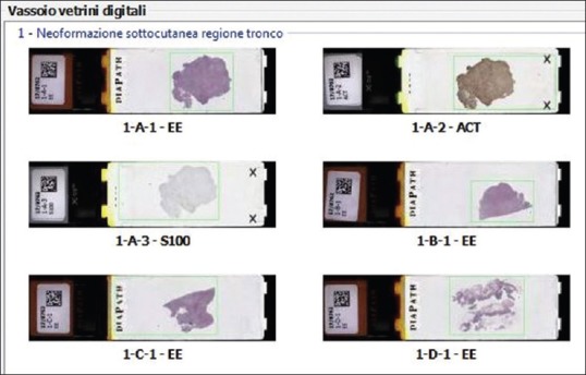
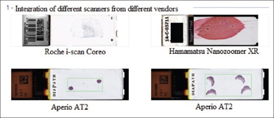
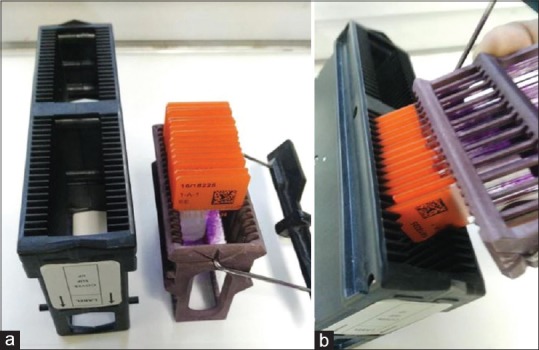
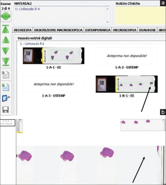
Methods: All (100%) permanent histopathology glass slides were digitized at ×20 using Aperio AT2 scanners. Compatible stain and scanning slide racks were employed to streamline operations. eSlide Manager software was bidirectionally interfaced with the anatomic pathology laboratory information system. Virtual slide trays connected to the two-dimensional (2D) barcode tracking system allowed pathologists to confirm that they were correctly assigned slides and that all tissues on these glass slides were scanned.
Results: Over 115,000 glass slides were digitized with a scan fail rate of around 1%. Drying glass slides before scanning minimized them sticking to scanner racks. Implementation required introduction of a 2D barcode tracking system and modification of histology workflow processes.
Conclusion: Our experience indicates that effective adoption of WSI for primary diagnostic use was more dependent on optimizing preimaging variables and integration with the laboratory information system than on information technology infrastructure and ensuring pathologist buy-in. Implementation of digital pathology for routine practice not only leveraged the benefits of digital imaging but also creates an opportunity for establishing standardization of workflow processes in the pathology laboratory.
Keywords: Digital pathology; informatics; pathology; whole slide imaging; workflow.
Conflict of interest statement
There are no conflicts of interest.
Figures
References
-
- Cheng CL, Tan PH. Digital pathology in the diagnostic setting: Beyond technology into best practice and service management. J Clin Pathol. 2017;70:454–7. - PubMed
-
- Ho J, Parwani AV, Jukic DM, Yagi Y, Anthony L, Gilbertson JR, et al. Use of whole slide imaging in surgical pathology quality assurance: Design and pilot validation studies. Hum Pathol. 2006;37:322–31. - PubMed
-
- Boyce BF. Whole slide imaging: Uses and limitations for surgical pathology and teaching. Biotech Histochem. 2015;90:321–30. - PubMed
-
- Cheng CL, Azhar R, Sng SH, Chua YQ, Hwang JS, Chin JP, et al. Enabling digital pathology in the diagnostic setting: Navigating through the implementation journey in an academic medical centre. J Clin Pathol. 2016;69:784–92. - PubMed
-
- Goacher E, Randell R, Williams B, Treanor D. The diagnostic concordance of whole slide imaging and light microscopy: A Systematic review. Arch Pathol Lab Med. 2017;141:151–61. - PubMed
LinkOut - more resources
Full Text Sources
Other Literature Sources
