. 2018 Jan 1;8(1):154-164.
eCollection 2018.

Isocorydine suppresses doxorubicin-induced epithelial-mesenchymal transition via inhibition of ERK signaling pathways in hepatocellular carcinoma

Affiliations

Isocorydine suppresses doxorubicin-induced epithelial-mesenchymal transition via inhibition of ERK signaling pathways in hepatocellular carcinoma

Jie-Xue Pan et al. Am J Cancer Res. .

Abstract

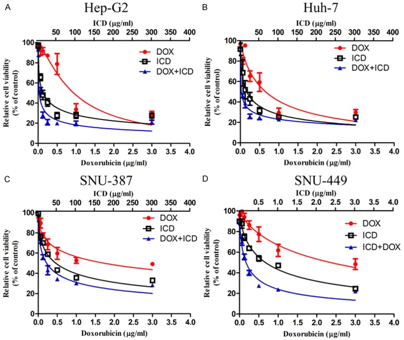
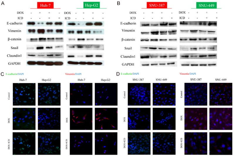
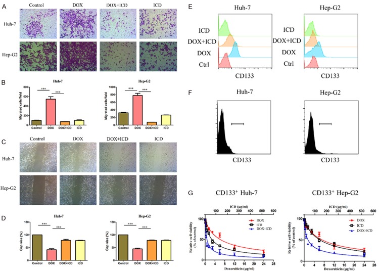
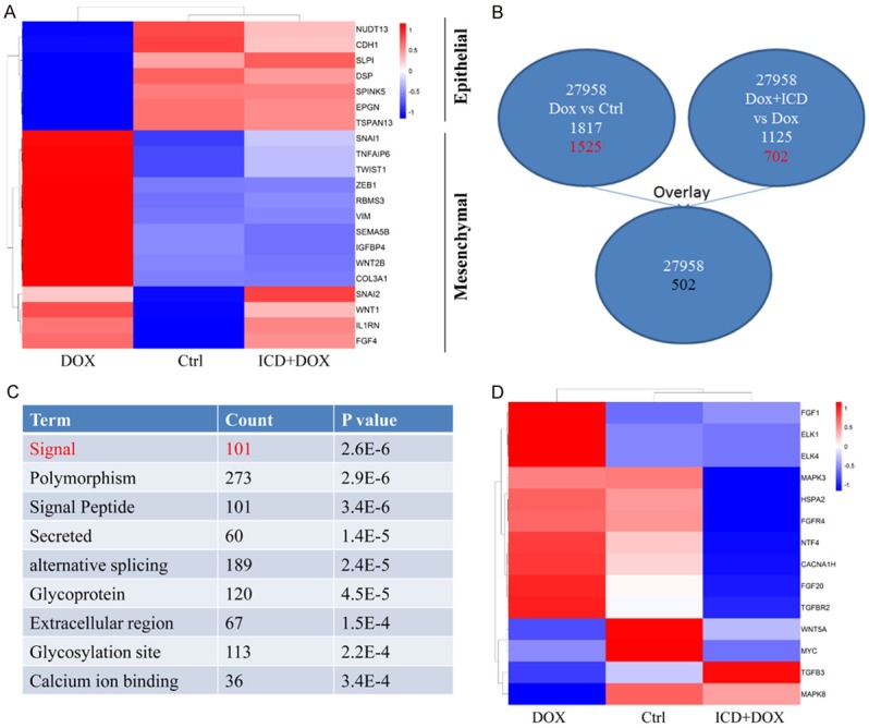
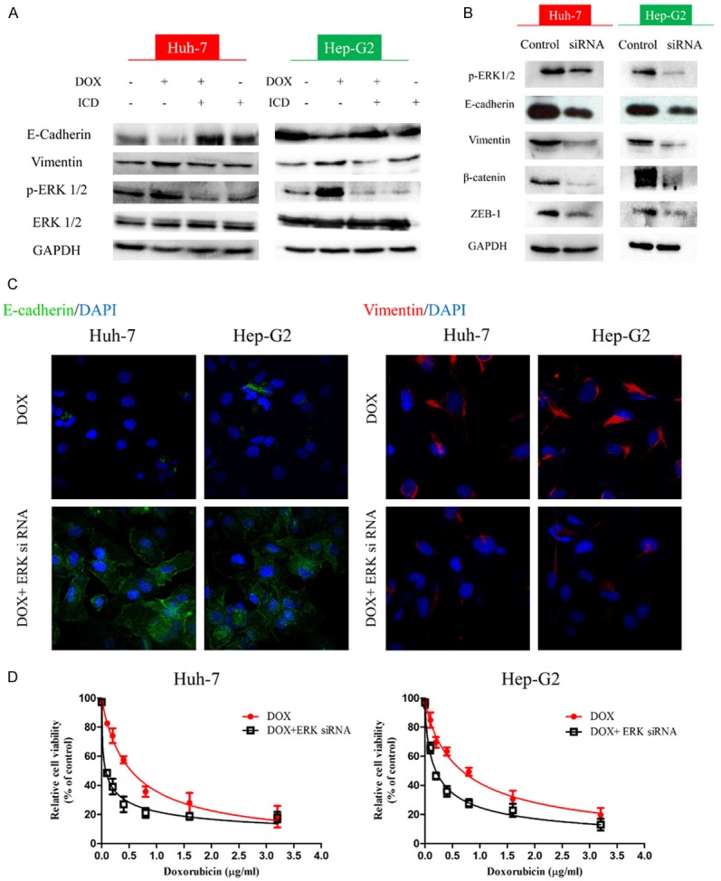
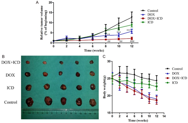
Doxorubicin (DOX) is a conventional and effective chemotherapeutic used in the treatment of hepatocellular carcinoma (HCC). However, doxorubicin administration may induce EMT, which results in the development of chemoresistance in HCC. Recent studies report that Isocorydine (ICD) selectively inhibits human cancer stem cells (CSCs), which have an important role in the development of chemoresistance. In this study, we observed that ICD co-administration enhanced DOX cytotoxicity in HCC cells, enabling the inhibition of DOX-induced epithelial-mesenchymal transition (EMT). Microarray data analysis revealed substantially decreased ERK signaling after ICD treatment. Additionally, we observed decreased IC50 for DOX upon ERK knockdown. Finally, we confirmed the enhanced efficacy of treatment with a combination of DOX and ICD in xenograft models. Collectively, the present study unveils the benefit of using DOX in combination with ICD for chemotherapy against HCC, revealing a novel potential anti-cancer strategy.

Keywords: Doxorubicin; EMT; HCC; Isocorydine.


Conflict of interest statement

None.

Figures

Figure 1
Figure 1
ICD enhances DOX inhibition of HCC proliferation in CCK-8 assay. Relative cell viability (mean ± SD) for ICD (black), DOX (red) and ICD + DOX (blue) in Huh-7, HepG2, SNU-449 and SNU-387 cells.
Figure 2
Figure 2
ICD alters the expression of DOX-induced EMT in HCC cells. Expression of EMT markers E-cadherin, Vimentin, β-catenin, Snail and Claundin1, as examined by western blot in Huh-7, Hep-G2 (A) and SNU-387, SNU-449 (B) cells, or treated with DOX, DOX + ICD, or ICD alone for 48 h. Immunofluorescence staining of E-cadherin and Vimentin in Huh-7, Hep-G2 (C) and SNU-387, SNU-449 (D) cells after DOX or DOX + ICD treatment (200x magnification).
Figure 3
Figure 3
ICD inhibits the DOX-induced tumor invasive capacity in HCC cells. (A) Transwell assays showing the invasion of Huh-7 and Hep-G2 cells for control, DOX, DOX + ICD or ICD group (100x magnification). (B) Number of cells per field that crossed the transwell chamber under the different treatment conditions (*P < 0.05, **P < 0.01, ***P < 0.001). (C) Wound healing assay for migration of Huh-7 and Hep-G2 cells in the control, DOX, DOX + ICD or ICD group (100x magnification). The broken white line indicates the wound areas without migrating cells. (D) The gap between the migrating cells is expressed as a percentage of the initial area. (E) Levels of CD133 in HCC cells (Huh-7 and Hep-G2), or HCC cells treated with DOX, DOX + ICD, or ICD alone for 48 h. Sorting CD133 + HCC cells (F) and the relative cell viability for ICD (black), DOX (red) and ICD + DOX (blue) in Huh-7 and Hep-G2 (G). One of three representative images are shown in (A, C).
Figure 4
Figure 4
MAPK cascade is operative in the ICD and DOX treated HCC cells. A. Heat map of the EMT-related genes in control, DOX and ICD + DOX samples. B. Schematic representation of comparing gene expression profiles in Huh-7 cells (2-fold difference, DOX vs. Ctrl; DOX + ICD vs. DOX) and overlay analysis. C. Hierarchical clustering of the above-mention genes. D. Heat map of the EMT-related genes in control, DOX and ICD + DOX.
Figure 5
Figure 5
ICD reverses DOX-induced EMT through ERK signaling. A. Expression of ERK1/2 examined by western blot in Huh-7, Hep-G2, or treated with DOX, DOX + ICD, or ICD alone for 48 h. B. Detection of EMT markers upon treatment with ERK1/2 siRNA. D. Immunofluorescence staining of E-cadherin and Vimentin in DOX and DOX + ERK siRNA in Huh-7 and Hep-G2 (200x magnification). C. The relative cell viability for DOX (red) and DOX + ERK siRNA (black) in Huh-7 and Hep-G2.
Figure 6
Figure 6
Effect of ICD, DOX, and their combination in nude mice implanted with subcutaneous HCC xenografts. A. Tumor volume over time in the control group (black), groups treated with DOX (blue), ICD (green) or DOX + ICD (red). Relative tumor volume ratios (% of original volume after initiation of therapy) are presented as the mean ± SD, (**P < 0.01, ***P < 0.001, for control vs. DOX group; ##P < 0.01, ###P < 0.001, for DOX + ICD vs. DOX alone). B. Resected tumors in the different groups. C. The body weight of mice after treatment.

References

    1. Chen W, Zheng R, Baade PD, Zhang S, Zeng H, Bray F, Jemal A, Yu XQ, He J. Cancer statistics in China, 2015. CA Cancer J Clin. 2016;66:115–132. - PubMed
    1. Ye LY, Chen W, Bai XL, Xu XY, Zhang Q, Xia XF, Sun X, Li GG, Hu QD, Fu QH, Liang TB. Hypoxia-Induced epithelial-to-mesenchymal transition in hepatocellular carcinoma induces an immunosuppressive tumor microenvironment to promote metastasis. Cancer Res. 2016;76:818–830. - PubMed
    1. Bruix J, Sherman M American Association for the Study of Liver Diseases. Management of hepatocellular carcinoma: an update. Hepatology. 2011;53:1020–1022. - PMC - PubMed
    1. Tang QH, Li AJ, Yang GM, Lai EC, Zhou WP, Jiang ZH, Lau WY, Wu MC. Surgical resection versus conformal radiotherapy combined with TACE for resectable hepatocellular carcinoma with portal vein tumor thrombus: a comparative study. World J Surg. 2013;37:1362–1370. - PubMed
    1. Moncharmont C, Levy A, Gilormini M, Bertrand G, Chargari C, Alphonse G, Ardail D, Rodriguez-Lafrasse C, Magne N. Targeting a cornerstone of radiation resistance: cancer stem cell. Cancer Lett. 2012;322:139–147. - PubMed

LinkOut - more resources