. 2017 Dec 18;7(2):e1378844.
doi: 10.1080/2162402X.2017.1378844. eCollection 2018.

Maturation of tertiary lymphoid structures and recurrence of stage II and III colorectal cancer

Affiliations

Maturation of tertiary lymphoid structures and recurrence of stage II and III colorectal cancer

Florian Posch et al. Oncoimmunology. .

Abstract

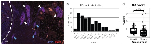
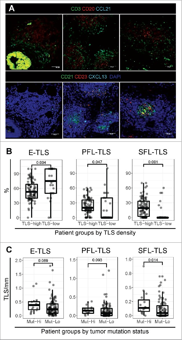
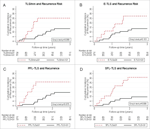
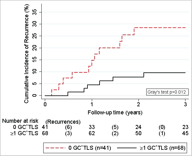
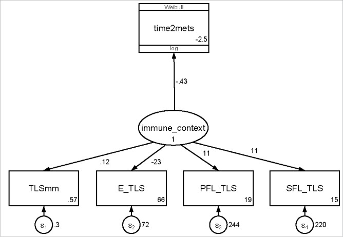
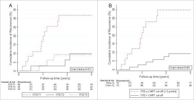
Tertiary lymphoid structures (TLS) are associated with favorable outcome in non-metastatic colorectal carcinoma (nmCRC), but the dynamics of TLS maturation and its association with effective anti-tumor immune surveillance in nmCRC are unclear. Here, we hypothesized that not only the number of TLS but also their composition harbors information on recurrence risk in nmCRC. In a comprehensive molecular, tissue, laboratory, and clinical analysis of 109 patients with stage II/III nmCRC, we assessed TLS numbers and degree of maturation in surgical specimens by multi-parameter immunofluorescence of follicular dendritic cell (FDC) and germinal center (GC) markers. TLS formed in most tumors and were significantly more prevalent in highly-microsatellite-instable (MSI-H) and/or BRAF-mutant nmCRC. We could distinguish three sequential TLS maturation stages which were characterized by increasing prevalence of FDCs and mature B-cells: [1] Early TLS, composed of dense lymphocytic aggregates without FDCs, [2] Primary follicle-like TLS, having FDCs but no GC reaction, and [3] Secondary follicle-like TLS, having an active GC reaction. A simple integrated TLS immunoscore reflecting these parameters identified a large subgroup of nmCRC patients with a very low risk of recurrence independently of clinical co-variables such as ECOG performance status, age, stage, and adjuvant chemotherapy. We conclude that (1) mismatch repair and BRAF mutation status are associated with the formation of TLS in nmCRC, (2) TLS formation in nmCRC follows sequential maturation steps, culminating in germinal center formation, and (3) this maturation process harbors important prognostic information on the risk of disease recurrence.

Keywords: Crohn's-like reaction; colorectal cancer; germinal center; immunoscore; recurrence; risk factor; structural equation model; tertiary lymphoid structures.


Figures

Figure 1.
Figure 1.
Tertiary lymphoid structures in stage II and III colorectal cancer. (A) The presence of TLS was analyzed in 109 CRC tissues by immunofluorescence and identified as dense B cell (CD20) aggregates. TLS near the invasive margin (white dashed line) of a CRC (T) and blood vessels (white stars). TLS with a central GC morphology (white arrowheads) or without (blue arrowheads) were counted. (B) The density of TLS in each patient was determined as the number of TLS per millimeter of tumor invasive front. (C) Comparison of TLS density in CRC patients with known MSI and BRAF mutation status. MSI-H and/or BRAF mutated CRCs (Mut-Hi, n = 15) were compared versus patients with wild type BRAF and MSS status (Mut-Lo, n = 94) by Wilcoxon rank-sum test.
Figure 2.
Figure 2.
Tertiary lymphoid structure maturation in stage II and III colorectal cancer. (A) The composition of TLS was analyzed by multi-parameter immunofluorescence using serial tissue sections of CRC. Two sets of antibodies were combined to visualize (1) the spatial organization of B cells (CD20), T cells (CD3), and CCL21 (top row), and (2) TLS maturation by the presence of FDCs (CD21), germinal center (GC) B cells (CD23), and CXCL13 (bottom row). (B) Different maturation stages were assessed by multiparameter immunofluorescence in CRC tissues. TLS in each maturation stage (E-TLS – dense lymphocytic clusters: CD21CD23, PFL-TLS – clusters with an FDC network: CD21+CD23, SFL-TLS – clusters with active GC reaction: CD21+CD23+) were counted for each patient and expressed as the proportion of the total TLS count. The proportions of the three maturation stages were compared in patients with high (n = 84) and low (n = 25) TLS density (cut-off at the 25th percentile of TLS density distribution (i.e. Q1)) by a Wilcoxon rank-sum test. (C) The density of each TLS maturation stage was compared for patients with mutated BRAF and/or MSI-H status (Mut-hi, n = 15) versus patients with wild type BRAF and MSS status (Mut-lo, n = 94) by a two-tailed Mann-Whitney U test.
Figure 3.
Figure 3.
Tertiary lymphoid structure parameters and 3-year risk of colorectal cancer recurrence. Patients with low TLS counts (A), high proportions of E-TLS (B), and low proportions of PFL-TLS (C) showed a tendency towards numerically higher risks of recurrence. Patients with low SFL-TLS proportion (D) experienced a significantly higher risk of recurrence. Absolute risk estimates corresponding to these curves are reported in Table 2. Recurrence risks were estimated with competing risk analysis, treating death-from-any-cause as the competing event of interest. In the risk table, numbers in brackets represent the number of recurrences in the respective time interval. Abbreviations: Q1 – Cut-off at the 25th percentile of the variables' distribution (i.e. quartile 1), Q3 – Cut-off at the 75th percentile of the variable's distribution (i.e. quartile 3).
Figure 4.
Figure 4.
Competing risk analysis of germinal-center-harboring TLS and 3-year risk of colorectal cancer recurrence. Cumulative incidences of recurrence were derived with competing risk analysis, accounting for death-from-any-cause as the competing risk of interest. Patients were dichotomized into two groups according to whether they had at least one TLS with an active GC reaction (n = 68) or not (n = 41). Risktables (“Number at risk”) report the number of patients in each group at risk of recurrence at the start of each time interval. The number of recurrences in each time interval (“Recurrences”) are reported in brackets.
Figure 5.
Figure 5.
Path diagram of the structural equation model for estimating the relationship between TLS parameters, a latent TLS immune contexture variable, and colorectal cancer recurrence. Numbers to the right of the path represent path coefficients (or the log(hazard ratio) for the path from “immune_context” to “time2mets”)). Numbers in the right bottom of the square boxes represent the intercepts of the path coefficients. Numbers adjacent to the round error terms represent the errors of the measurements. Abbreviations: TLSmm – TLS count, E-TLS – Early TLS proportion, PFL-TLS – primary follicle like TLS proportion, SFL-TLS – secondary follicle like TLS proportion.
Figure 6.
Figure 6.
Competing risk analysis of the Integrated TLS ImmunoScore (ITIS) and 3-year risk of colorectal cancer recurrence. Cumulative incidences of recurrence were derived with competing risk analysis, accounting for death-from-any-cause as the competing risk of interest. (A) The ITIS was categorized empirically into tertiles (T1, T2, and T3). (B) The ITIS was dichotomized into a binary variable at the cut-off suggested by the Classification And Regression Tree (CART) analysis at -12 points. Risktables (“Number at risk”) report the number of patients in each group at risk of recurrence at the start of each time interval. The number of recurrences in each time interval (“Recurrences”) are reported in brackets.

References

    1. Siegel RL, Miller KD, Jemal A. Cancer Statistics, 2017. CA Cancer J Clin. 2017;67:7-30. doi:10.3322/caac.21387. PMID:28055103 - DOI - PubMed
    1. Torre LA, Siegel RL, Ward EM, Jemal A. Global Cancer Incidence and Mortality Rates and Trends–An Update. Cancer Epidemiol Biomarkers Prev. 2016;25:16-27. doi:10.1158/1055-9965.EPI-15-0578. PMID:26667886 - DOI - PubMed
    1. Brenner H, Kloor M, Pox CP. Colorectal cancer. Lancet (London, England). 2014;383:1490-502. doi:10.1016/S0140-6736(13)61649-9. PMID:24225001 - DOI - PubMed
    1. Lech G, Slotwinski R, Slodkowski M, Krasnodebski IW. Colorectal cancer tumour markers and biomarkers: Recent therapeutic advances. World J Gastroenterol. 2016;22:1745-55. doi:10.3748/wjg.v22.i5.1745. PMID:26855534 - DOI - PMC - PubMed
    1. Galon J, Mlecnik B, Bindea G, Angell HK, Berger A, Lagorce C, Lugli A, Zlobec I, Hartmann A, Bifulco C, et al.. Towards the introduction of the ‘Immunoscore’ in the classification of malignant tumours. J Pathol. 2014;232:199-209. doi:10.1002/path.4287. PMID:24122236 - DOI - PMC - PubMed

Publication types

LinkOut - more resources