Review
. 2018 Jan 24:6:1.
doi: 10.3389/fcell.2018.00001. eCollection 2018.

Subversion of the Endocytic and Secretory Pathways by Bacterial Effector Proteins

Affiliations
Review

Subversion of the Endocytic and Secretory Pathways by Bacterial Effector Proteins

Mary M Weber et al. Front Cell Dev Biol. .

Abstract

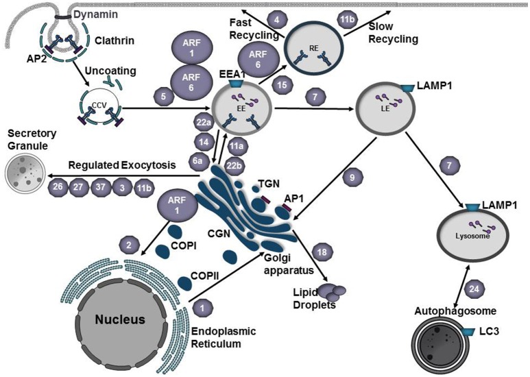
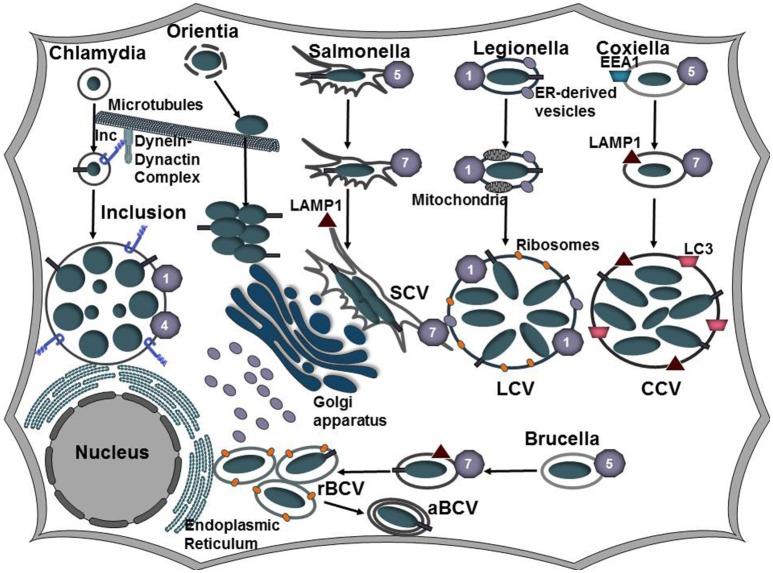
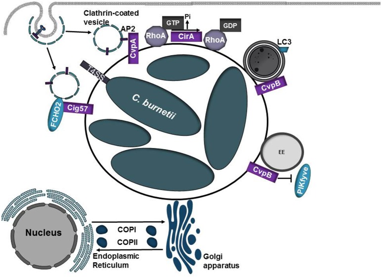
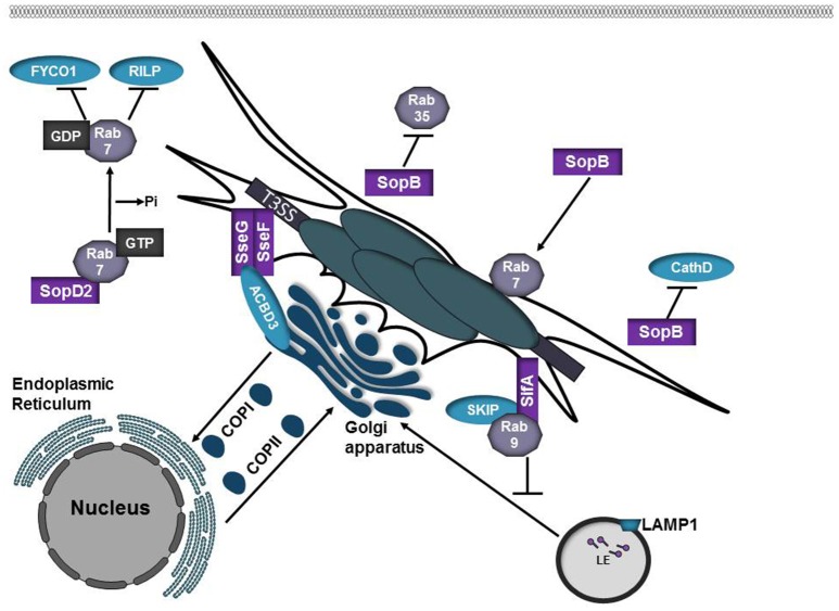
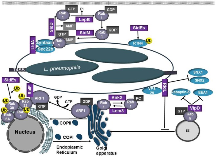
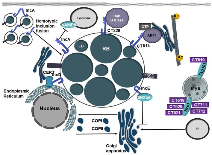
Intracellular bacteria have developed numerous strategies to hijack host vesicular trafficking pathways to form their unique replicative niches. To promote intracellular replication, the bacteria must interact with host organelles and modulate host signaling pathways to acquire nutrients and membrane for the growing parasitophorous vacuole all while suppressing activation of the immune response. To facilitate host cell subversion, bacterial pathogens use specialized secretion systems to deliver bacterial virulence factors, termed effectors, into the host cell that mimic, agonize, and/or antagonize the function of host proteins. In this review we will discuss how bacterial effector proteins from Coxiella burnetii, Brucella abortus, Salmonella enterica serovar Typhimurium, Legionella pneumophila, Chlamydia trachomatis, and Orientia tsutsugamushi manipulate the endocytic and secretory pathways. Understanding how bacterial effector proteins manipulate host processes not only gives us keen insight into bacterial pathogenesis, but also enhances our understanding of how eukaryotic membrane trafficking is regulated.

Keywords: Brucella; Chlamydia; Coxiella; Legionella; Orientia; Salmonella; secreted effector; vesicle trafficking.


Figures

Figure 1
Figure 1
Vesicle trafficking pathways of the endocytic and secretory pathways. Small GTPases of the Arf family recruit protein coats to the donor membrane to initiate vesicle budding. Clathrin coated vesicles are generated at the plasma membrane or trans-Golgi network (TGN) and mediate cargo transport to endosomes. Adaptor proteins, such as AP1 and AP3, recognize signal sequences in the C-terminal tails of transmembrane proteins and participate in cargo selection. Coat complex protein I (COPI) and coat complex protein II (COPII) mediate transport from the Golgi to the endoplasmic reticulum (ER) or from the ER to the Golgi, respectively. At the plasma membrane, vesicle scission is mediated by dynamin and vesicles are subsequently transported to their destination along cytoskeletal tracks. Rab GTPases (gray hexagons) play a key role in regulating vesicle transport along the endocytic and secretory pathways. Rab5 mediates fusion of endocytic vesicles to form the early endosome (EE). Maturation into a late endosome (LE) requires Rab conversion from Rab5 to Rab7. Cargo destined for degradation undergoes fusion with lysosomes. Rab24 mediates formation of autophagosomes, which can subsequently fuse with lysosomes to create autolysosomes. Traffic between EE and recycling endosome (RE) is mediated by Rab15. Rab4 and Rab11b regulate fast and slow recycling, respectively. Rab1 mediates ER-Golgi traffic whereas Rab2 regulates retrograde traffic from the Golgi-ER. Regulated exocytosis by secretory granules involves Rab26, Rab27, Rab37, Rab3, or Rab11b. Transport between the EE and Golgi or the LE and Golgi is mediated by Rab22a, Rab14, Rab6a, or Rab9, respectively. Cargo from the TGN is transported to the plasma membrane via fusion with EEs and this process is mediated by Rab22b or Rab11a. Rab18 regulates the formation of lipid droplets. EE early endosome, LE late endosome, RE recycling endosome TGN trans-Golgi Network, CGN cis-Golgi Network, COPI coat protein I, COPII coat protein II, CCV clathrin-coated vesicle, AP1 adaptor protein 1, AP2 adaptor protein 2.
Figure 2
Figure 2
Establishing a replicative niche. Following uptake by a host cell, intracellular bacteria manipulate the endocytic and secretory pathways of the host cell to establish a replicative niche. Coxiella, Salmonella, and Brucella associate with the endocytic pathways as evident by Rab5, Rab7, and LAMP1 that decorate the Coxiella-containing vacuole (CCV), Salmonella-containing vacuole (SCV), and Brucella-containing vacuole (BCV), respectively. As the CCV matures, it undergoes fusions with autophagosomes and acquires LC3. Vacuolar acidification induces expression of the C. burnetii Dot/Icm and the Brucella VirB type IV secretion systems. Following transient vacuolar acidification, the BCV is redirected to and fuses with the endoplasmic reticulum to form the rBCV. Some bacteria undergo interactions with autophagosomes (aBCV) as a possible exit mechanism. Salmonella replicates in the peri-Golgi region and induces the formation of tubular membranes, referred to as Salmonella-induced filaments (SIFs) that are enriched in LAMP1. L. pneumophila secretes Dot/Icm effector proteins to bypass the endocytic pathway and instead redirects ER-derived vesicles to the LCV. C. trachomatis also evades the endocytic pathway and inhibits fusions with lysosomes. Modification of the inclusion membrane by incorporation of type III secreted effector proteins, termed inclusion membrane proteins (Incs), promotes trafficking of the inclusion along microtubules to the microtubule organizing center (MTOC). The bacteria undergo conversion from the elementary body (EB) to the replicative body (RB) and the bacteria replicate by binary fission. Following internalization, Orientia tsutsugamushi escapes the host-derived vacuole and traffics along microtubules to replicate in the cytosol juxtaposed to the ER and Golgi apparatus.
Figure 3
Figure 3
Diverse roles of C. burnetii T4SS effector proteins. Coxiella burnetii replicates in an acidic phagolysosome-like compartment termed the Coxiella-containing vacuole (CCV). Acidification of the CCV promotes activation of the Dot/Icm type IV secretion system (T4SS) which translocates over 130 proteins into the eukaryotic host cell. As the CCV matures, it undergoes fusions with autophagosomes and acquires the autophagosomal maker LC3. CvpA and Cig57 modulate clathrin-dependent vesicular transport pathways through interactions with adaptor protein 2 (AP2) or FCHO2, respectively. CirA acts as a GTPase activating protein (GAP) for RhoA. CvpB/Cig2 promotes CCV expansion by targeting endosomes and autophagosomes. CvpB/Cig2 interferes with PI 5-kinase PIKfyve access to early endosomes, resulting in increased levels of PI(3)P on CCVs.
Figure 4
Figure 4
Salmonella SPI-2 effector proteins participate in SCV formation. SopB is phosphoinositide phosphatase that alters the membrane surface charge of the SCV. This prevents targeting by Rab35 while promoting recruitment of Rab7. SopB also inhibits targeting of the lysosomal protease Cathepsin D to the SCV. SopD2 is a Rab7 GAP. Inactivation of Rab7 by SopD2 prevents interactions with the Rab7 effectors RILP and FYCO1. SifA is inserted into the SCV membrane where it interacts with SKIP/PLEKHM2. SifA-SKIP sequesters Rab9 at the SCV, inhibiting retrograde trafficking of mannose-phosphate receptors (MPRs) to the trans-Golgi network. SseF and SseG are integral membrane effector proteins that form a trimolecular complex with ACBD3 to anchor the SCV to the Golgi apparatus.
Figure 5
Figure 5
Role of L. pneumophilia T4SS effector proteins in modulation of the endocytic and secretory pathways. L. pneumophila recruits the small GTPases Rab1 and Arf1 to the LCV membrane to co-opt ER-Golgi vesicle trafficking. RalF and SidM/DrrA are Arf1 and Rab1 GEFs, respectively. SidM/DrrA can covalently modify Rab1 through AMPylation whereas SidD functions as a deAMPylase to remove the AMP residue, making Rab1 susceptible to inactivation by the bacterial GAP LepB. Activated Rab1 promotes binding of LidA, which cooperates with SidM/DrrA to promote tethering and fusion between the LCV and ER-derived vesicles. AnkX and Lem3 modify Rab1 activity through phosphorylcholination (PCylation) or by removal of the phosphorylcholine moiety, respectively. The SidE family of T4SS effectors (SidE, SdeA, SdeB, SdeC) ubiquitinate Rab1, Rab6, Rab30, and Rab33b. SidE effectors also ubiquitinate reticulon 4 (RTN4) to induce ER rearrangements and promote RTN4 recruitment to the LCV. RidL binds PtdIns(3)P and the retromer subunit VPS29, inhibiting SNX 1 and 2 binding to the LCV. VipD prevents endosomal maturation by binding GTP-bound Rab5 and Rab22, inhibiting subsequent interactions with the Rab effectors Rabaptin-5 and EEA1.
Figure 6
Figure 6
Inclusion membrane proteins and secreted effectors from C. trachomatis co-opt host vesicular trafficking pathways to promote inclusion development. Chlamydia trachomatis replicates in a parasitophorous vacuole, termed the inclusion. Early in the infection cycle the inclusion is modified through the incorporation of type III secreted effector proteins known as inclusion membrane proteins (Incs). IncA possesses 2 coiled-coil domains that are homologous to eukaryotic SNARE motifs and these domains are required to mediate homotypic fusion of inclusions. IncA is also able to interact with VAMP8 on lysosomes and may prevent inclusion fusion with lysosomes. C. trachomatis manipulates and recruits Rab and Arf GTPases to the inclusion membrane through interactions with the inclusion membrane proteins CT229 and CT813, respectively. While the mechanism of CT229-Rab interactions is ill-defined, CT813 is presumed to modulate Arf GTPases to control Golgi ministack positioning by regulating microtubule posttranslational modification. IncE interacts with SNX5/6, components of the retromer complex, to disrupt trafficking to the trans-Golgi network and to promote chlamydial infection. To acquire the sphingomyelin precursor ceramide, IncD interacts with the Pleckstrin homology (PH) domain of CERT allowing ceramide to be directly transferred from the ER to the inclusion membrane. C. trachomatis also secretes a large repertoire of type III proteins into the eukaryotic cells. CT619, CT620, CT621, CT711, CT712 are type III substrates that interact with Hrs and/or Tsg101, components of the ESCRT pathway.

References

    1. Abrahams G. L., Müller P., Hensel M. (2006). Functional dissection of SseF, a type III effector protein involved in positioning the Salmonella-containing vacuole. Traffic 7, 950–965. 10.1111/j.1600-0854.2006.00454.x - DOI - PubMed
    1. Aeberhard L., Banhart S., Fischer M., Jehmlich N., Rose L., Koch S., et al. . (2015). The proteome of the isolated Chlamydia trachomatis containing vacuole reveals a complex trafficking platform enriched for retromer components. PLoS Pathog. 11:e1004883. 10.1371/journal.ppat.1004883 - DOI - PMC - PubMed
    1. Allgood S. C., Duenas B. P. R., Noll R. R., Pike C., Lein S., Neunuebel M. R. (2017). Legionella effector AnkX disrupts host cell endocytic recycling in a phosphocholination-dependent manner. Front. Cell Inf. Microbiol. 7:397. 10.3389/fcimb.2017.00397 - DOI - PMC - PubMed
    1. Al-Zeer M. A., Al-Younes H. M., Kerr M., Abu-Lubad M., Gonzalez E., Brinkmann V., et al. . (2014). Chlamydia trachomatis remodels stable microtubules to coordinate Golgi stack recruitment to the chlamydial inclusion surface. Mol. Microbiol. 94, 1285–1297. 10.1111/mmi.12829 - DOI - PubMed
    1. Anderson C. J., Kendall M. M. (2017). Salmonella enterica serovar Typhimurium strategies for host adaptation. Front. Microbiol. 8:1983 10.3389/fmicb.2017.01983 - DOI - PMC - PubMed

LinkOut - more resources