. 2018 Aug;75(15):2843-2856.
doi: 10.1007/s00018-018-2764-5. Epub 2018 Feb 7.

Stem cells from human apical papilla decrease neuro-inflammation and stimulate oligodendrocyte progenitor differentiation via activin-A secretion

Affiliations

Stem cells from human apical papilla decrease neuro-inflammation and stimulate oligodendrocyte progenitor differentiation via activin-A secretion

Pauline De Berdt et al. Cell Mol Life Sci. 2018 Aug.

Erratum in

Abstract

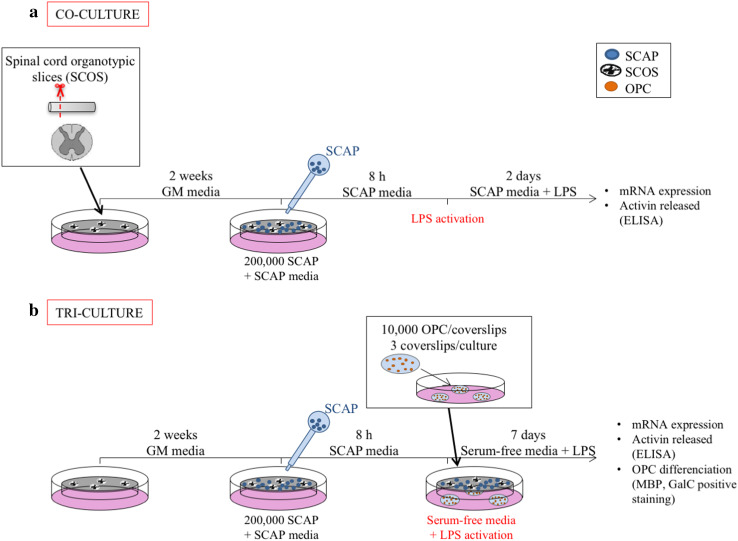
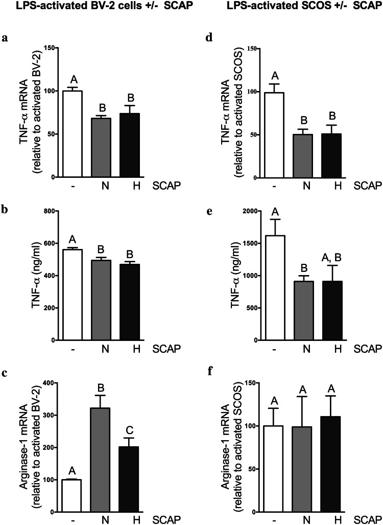
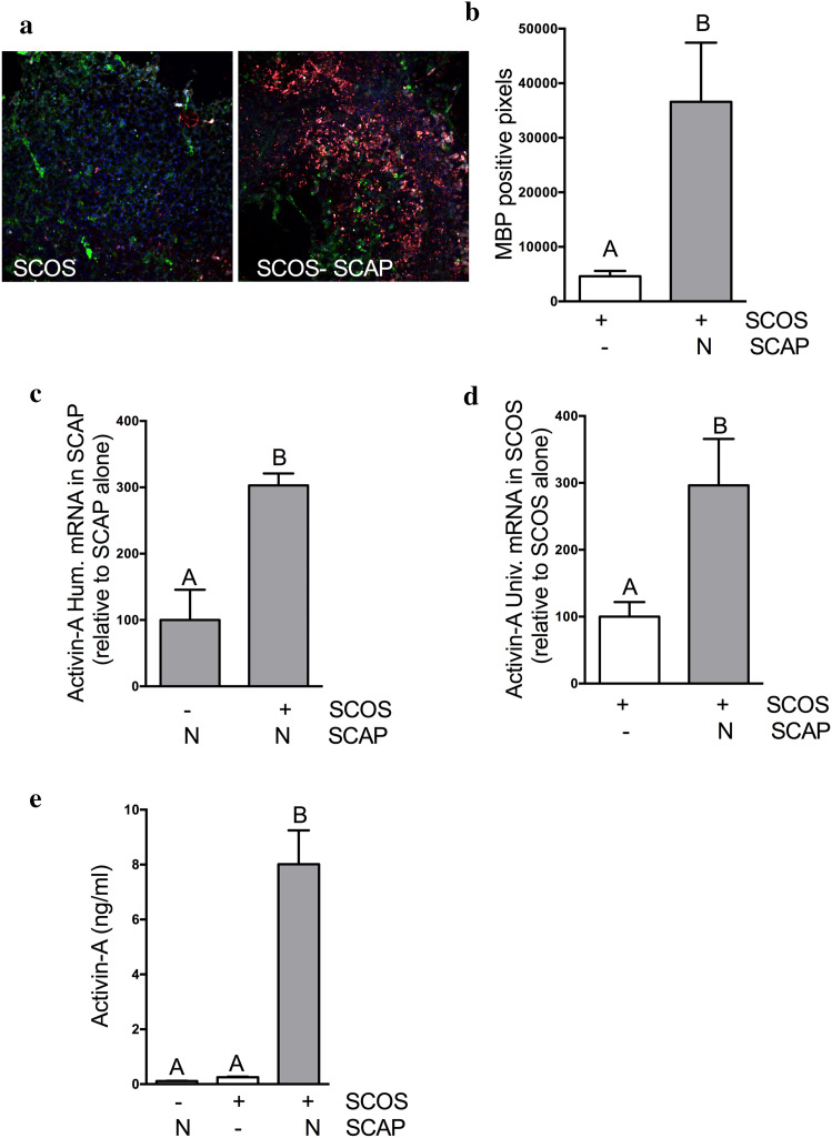
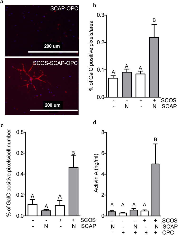
Secondary damage following spinal cord injury leads to non-reversible lesions and hampering of the reparative process. The local production of pro-inflammatory cytokines such as TNF-α can exacerbate these events. Oligodendrocyte death also occurs, followed by progressive demyelination leading to significant tissue degeneration. Dental stem cells from human apical papilla (SCAP) can be easily obtained at the removal of an adult immature tooth. This offers a minimally invasive approach to re-use this tissue as a source of stem cells, as compared to biopsying neural tissue from a patient with a spinal cord injury. We assessed the potential of SCAP to exert neuroprotective effects by investigating two possible modes of action: modulation of neuro-inflammation and oligodendrocyte progenitor cell (OPC) differentiation. SCAP were co-cultured with LPS-activated microglia, LPS-activated rat spinal cord organotypic sections (SCOS), and LPS-activated co-cultures of SCOS and spinal cord adult OPC. We showed for the first time that SCAP can induce a reduction of TNF-α expression and secretion in inflamed spinal cord tissues and can stimulate OPC differentiation via activin-A secretion. This work underlines the potential therapeutic benefits of SCAP for spinal cord injury repair.

Keywords: Dental stem cells; Differentiation; Inflammation; Oligodendrocyte progenitor cells; Spinal cord.


Figures

Fig. 1
Fig. 1
Graphical experimental plan. a Co-culture model of SCOS with SCAP on insert. b Tri-culture model of SCOS with SCAP on inserts and with OPC isolated from adult rat spinal cord seeded on coverslips. Groups without SCAP are presented in white, groups with SCAP grown in normoxia (N) in light grey and groups with SCAP grown in hypoxia (H) in dark grey
Fig. 2
Fig. 2
SCAP modulate the inflammatory markers gene expression and secretion. ac SCAP impact on LPS-activated BV-2 cells, 48 h after incubation. a SCAP impact on TNF-α gene expression of BV-2 cells evaluated by RT-qPCR (N = 2, n = 4). b Impact of SCAP co-cultured with BV-2 cells on BV-2 cells TNF-α production in culture media (ELISA) (N = 2, n = 4). c SCAP impact on arginase-1 gene expression of BV-2 cells (N = 2, n = 4). df SCAP impact on LPS-activated SCOS, 48 h after incubation. d Impact of SCAP on SCOS TNF-α gene expression (N = 2, n = 4). E: Impact of SCAP co-cultured with SCOS on TNF-α production in culture media (ELISA) (N = 2, n = 4). f Impact of SCAP on SCOS arginase gene expression (N = 2, n = 4). Conditions not related by the same letter are significantly different. Groups without SCAP are presented in white, groups with SCAP grown in normoxia (N) in light grey and groups with SCAP grown in hypoxia (H) in dark grey
Fig. 3
Fig. 3
SCOS–SCAP co-culture increased the expression of MBP in SCOS and induces activin-A secretion. a OPC were identified in SCOS by immunofluorescence using NG2 (green) staining and oligodendrocytes were identified using MBP (red) and CC1 (white) staining. b MBP positive pixels were quantified in SCOS cultured with and without SCAP (N = 2, n = 3). c Activin-A gene in SCAP 48 h after incubation with SCOS (N = 2, n = 3). d Activin-A gene expression in SCOS 48 h after incubation with SCAP (N = 2, n = 3). e Activin-A quantification in culture media using ELISA (N = 3, n = 3). Conditions not related by the same letter are significantly different. Groups without SCAP are presented in white, groups with SCAP grown in normoxia (N) in light grey and groups with SCAP grown in hypoxia (H) in dark grey
Fig. 4
Fig. 4
SCOS–SCAP promotes adult OPC differentiation and induces activin-A secretion. a Oligodendrocytes were identified by immunofluorescence using GalC staining 7 days after incubation of adult rat spinal cord OPC with SCOS and SCAP. bc GalC positive pixels were quantified by ImageJ software  per area (b) and cell numer (c) [N = 2, n = 3 (3 pictures/coverslips)]. d Activin-A quantification in culture media after 48 h of incubation (ELISA) (N = 3, n = 3). Conditions not related by the same letter are significantly different. Groups without SCAP are presented in white, groups with SCAP grown in normoxia (N) in light grey and groups with SCAP grown in hypoxia (H) in dark grey
Fig. 5
Fig. 5
Activin-A produced by SCAP and SCOS induces OPC differentiation. a Activin-A quantification in culture media (ELISA) 48 h after incubation (N = 2, n = 3). b Oligodendrocytes were identified by immunofluorescence using GalC staining 7 days after incubation. cd GalC positive pixels were quantified by ImageJ software per area (c) and cell number (d) [N = 2, n = 3 (3 pictures/coverslips)]. Conditions not related by the same letter are significantly different. Groups without SCAP are presented in white, groups with SCAP grown in normoxia (N) in light grey and groups with SCAP grown in hypoxia (H) in dark grey

Similar articles

Cited by

References

    1. Bianco J, De Berdt P, Deumens R, des Rieux A. Taking a bite out of spinal cord injury: do dental stem cells have the teeth for it? Cell Mol Life Sci. 2016;73(7):1413–1437. doi: 10.1007/s00018-015-2126-5. - DOI - PMC - PubMed
    1. Hall ED, Springer JE. Neuroprotection and acute spinal cord injury: a reappraisal. NeuroRx. 2004;1(1):80–100. doi: 10.1602/neurorx.1.1.80. - DOI - PMC - PubMed
    1. Ekdahl CT, Claasen JH, Bonde S, Kokaia Z, Lindvall O. Inflammation is detrimental for neurogenesis in adult brain. Proc Natl Acad Sci USA. 2003;100(23):13632–13637. doi: 10.1073/pnas.2234031100. - DOI - PMC - PubMed
    1. Fitch MT, Silver J. CNS injury, glial scars, and inflammation: inhibitory extracellular matrices and regeneration failure. Exp Neurol. 2008;209(2):294–301. doi: 10.1016/j.expneurol.2007.05.014. - DOI - PMC - PubMed
    1. Kyritsis N, Kizil C, Brand M. Neuroinflammation and central nervous system regeneration in vertebrates. Trends Cell Biol. 2014;24(2):128–135. doi: 10.1016/j.tcb.2013.08.004. - DOI - PubMed

MeSH terms

LinkOut - more resources