Review
. 2018 Feb 1;10(2):a023200.
doi: 10.1101/cshperspect.a023200.

Muscle Contraction

Affiliations
Review

Muscle Contraction

H Lee Sweeney et al. Cold Spring Harb Perspect Biol. .

Abstract

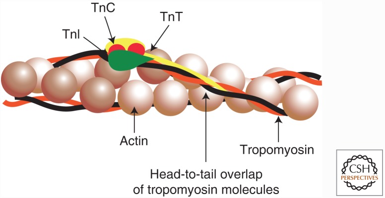
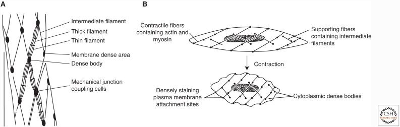
Muscle cells are designed to generate force and movement. There are three types of mammalian muscles-skeletal, cardiac, and smooth. Skeletal muscles are attached to bones and move them relative to each other. Cardiac muscle comprises the heart, which pumps blood through the vasculature. Skeletal and cardiac muscles are known as striated muscles, because the filaments of actin and myosin that power their contraction are organized into repeating arrays, called sarcomeres, that have a striated microscopic appearance. Smooth muscle does not contain sarcomeres but uses the contraction of filaments of actin and myosin to constrict blood vessels and move the contents of hollow organs in the body. Here, we review the principal molecular organization of the three types of muscle and their contractile regulation through signaling mechanisms and discuss their major structural and functional similarities that hint at the possible evolutionary relationships between the cell types.


Figures

Figure 1.
Figure 1.
Organization of the sarcomere of skeletal muscle. Electron micrograph of a longitudinal thin section through a muscle fiber, with the fiber long axis horizontal. Two complete sarcomeres are shown, and elements of the sarcoplasmic reticulum separate myofibrils in the longitudinal direction. The major bands and lines are indicated, notably the thin, distinctive Z-line flanked by two low-density half I-bands, with very dense A-bands containing the thick filaments. The relative densities of the bands in this electron micrograph are related to their content of protein. The light band on either side of the M-line shows the extent of the cross-bridge-free regions of the thick filaments. The fine transverse periodicity across the A-band, at ∼43 nm, is due to the periodic structure of the thick filament backbone, which in turn determines the position of relaxed cross-bridges on the surface of the filaments and is enhanced by the presence of accessory proteins.
Figure 2.
Figure 2.
Regulation of thin filaments. The thin filament proteins tropomyosin and troponin regulate the contraction of striated muscle. Tropomyosin is an elongated, coiled-coil molecule that binds to the surface of the thin actin filament. Troponin comprises three subunits—troponin T (TnT), which binds to tropomyosin; troponin I (TnI), which binds to both actin and tropomyosin; and troponin C (TnC), which confers Ca2+ sensitivity to the system. The relative position of these proteins on the actin filament is schematized.
Figure 3.
Figure 3.
The myosin molecule and its proteolytic fragments. Illustrated is the myosin molecule of muscle—myosin II—and the fragments that can be generated by limited proteolysis. The catalytic portion of the molecule is in the two enzymatically functional head (S1) fragments, each containing a nucleotide-binding site and an actin-binding site. The S1 fragment is commonly referred to as the “myosin head.” The remainder of the myosin molecule—the “rod”—comprising the amino-terminal portion (S2) and light meromyosin (LMM) fragments, forms an α-helical coiled coil. LMM forms the backbone of myosin filaments. A two-headed, catalytically active fragment of myosin known as heavy meromyosin (HMM) does not form filaments, and this serves a useful purpose for solution-biochemistry studies.
Figure 4.
Figure 4.
The contractile apparatus of smooth muscle. (A) Schematic of the key components of the force-generating protein network in mammalian smooth muscle. (B) The organization and rearrangements of the smooth muscle cell cytoskeleton during contraction.

References

    1. Almenar-Queralt A, Lee A, Conley CA, Ribas de Pouplana L, Fowler VM. 1999. Identification of a novel tropomodulin isoform, skeletal tropomodulin, that caps actin filament pointed ends in fast skeletal muscle. J Biol Chem 274: 28466–28475. - PubMed
    1. Bang ML, Centner T, Fornoff F, Geach AJ, Gotthardt M, McNabb M, Witt CC, Labeit D, Gregorio CC, Granzier H, et al. 2001. The complete gene sequence of titin, expression of an unusual approximately 700-kDa titin isoform, and its interaction with obscurin identify a novel Z-line to I-band linking system. Circ Res 89: 1065–1072. - PubMed
    1. Bers DM. 2002. Cardiac excitation-contraction coupling. Nature 415: 198–205. - PubMed
    1. Bresnick AR. 1999. Molecular mechanisms of nonmuscle myosin-II regulation. Curr Opin Cell Biol 11: 26–33. - PubMed
    1. Brooke MH, Kaiser KK. 1970. Muscle fiber types: How many and what kind? Arch Neurol 23: 369–379. - PubMed

LinkOut - more resources