

Recent Advances of Malaria Parasites Detection Systems Based on Mathematical Morphology
- PMID: 29419781
- PMCID: PMC5856187
- DOI: 10.3390/s18020513
Recent Advances of Malaria Parasites Detection Systems Based on Mathematical Morphology
Abstract
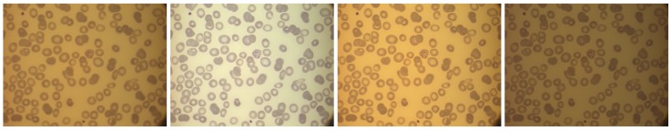
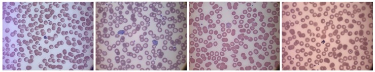
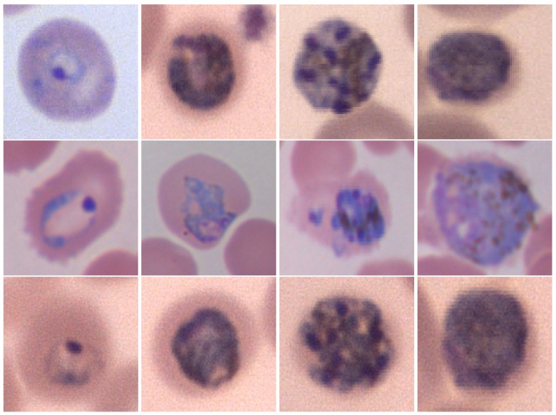
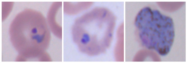
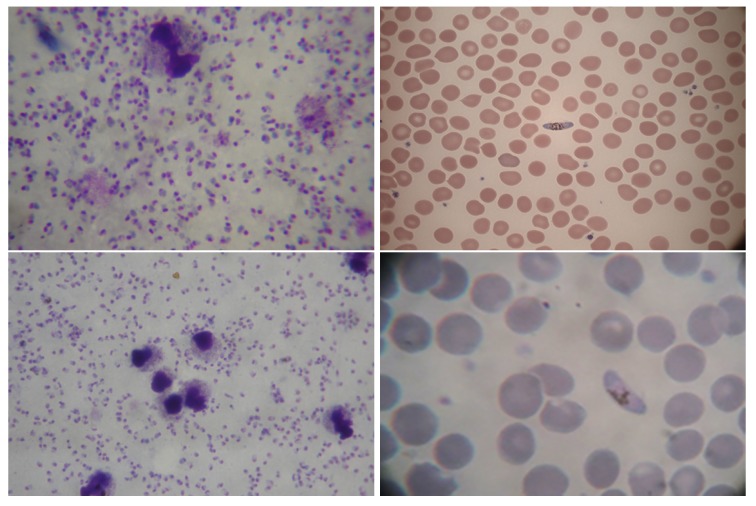
Malaria is an epidemic health disease and a rapid, accurate diagnosis is necessary for proper intervention. Generally, pathologists visually examine blood stained slides for malaria diagnosis. Nevertheless, this kind of visual inspection is subjective, error-prone and time-consuming. In order to overcome the issues, numerous methods of automatic malaria diagnosis have been proposed so far. In particular, many researchers have used mathematical morphology as a powerful tool for computer aided malaria detection and classification. Mathematical morphology is not only a theory for the analysis of spatial structures, but also a very powerful technique widely used for image processing purposes and employed successfully in biomedical image analysis, especially in preprocessing and segmentation tasks. Microscopic image analysis and particularly malaria detection and classification can greatly benefit from the use of morphological operators. The aim of this paper is to present a review of recent mathematical morphology based methods for malaria parasite detection and identification in stained blood smears images.
Keywords: malaria; mathematical morphology; medical image analysis; red blood cells segmentation.
Conflict of interest statement
The authors declare no conflict of interest.
Figures
References
-
- Loddo A., Putzu L., Di Ruberto C., Fenu G. A Computer-Aided System for Differential Count from Peripheral Blood Cell Images; Proceedings of the 2016 12th International Conference on Signal-Image Technology Internet-Based Systems (SITIS); Naples, Italy. 28 November–1 December 2016; pp. 112–118.
-
- Di Ruberto C., Loddo A., Putzu L. A leucocytes count system from blood smear images: Segmentation and counting of white blood cells based on learning by sampling. Mach. Vis. Appl. 2016;27:1151–1160. doi: 10.1007/s00138-016-0812-4. - DOI
-
- World Health Organization (WHO) Malaria Fact Sheet December 2016. [(accessed on 6 March 2017)]; Available online: http://www.who.int/mediacentre/factsheets/fs094/en/
-
- Somasekar J. Computer vision for malaria parasite classification in erythrocytes. Int. J. Comput. Sci. Eng. 2011;3:2251–2256.
-
- Soille P. Morphological Image Analysis: Principles and Applications. Springer; Berlin, Germany: 2004. p. 392.
Publication types
MeSH terms
LinkOut - more resources
Full Text Sources
Other Literature Sources
Medical
