. 2018 Apr;12(4):514-528.
doi: 10.1002/1878-0261.12178. Epub 2018 Feb 20.

STAT1 is a sex-specific tumor suppressor in colitis-associated colorectal cancer

Affiliations

STAT1 is a sex-specific tumor suppressor in colitis-associated colorectal cancer

Ilija Crnčec et al. Mol Oncol. 2018 Apr.

Abstract

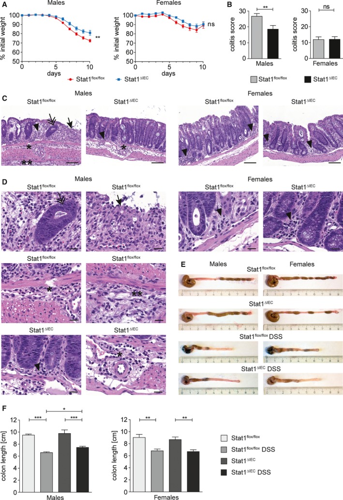
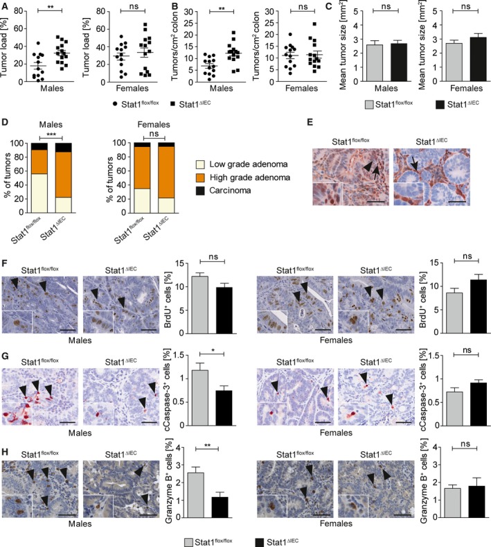
The interferon-inducible transcription factor STAT1 is a tumor suppressor in various malignancies. We investigated sex-specific STAT1 functions in colitis and colitis-associated colorectal cancer (CRC) using mice with specific STAT1 deletion in intestinal epithelial cells (STAT1∆IEC ). Male but not female STAT1∆IEC mice were more resistant to DSS-induced colitis than sex-matched STAT1flox/flox controls and displayed reduced intraepithelial infiltration of CD8+ TCRαβ+ granzyme B+ T cells. Moreover, DSS treatment failed to induce expression of T-cell-attracting chemokines in intestinal epithelial cells of male but not of female STAT1∆IEC mice. Application of the AOM-DSS protocol for induction of colitis-associated CRC resulted in increased intestinal tumor load in male but not in female STAT1∆IEC mice. A sex-specific stratification of human CRC patients corroborated the data obtained in mice and revealed that reduced tumor cell-intrinsic nuclear STAT1 protein expression is a poor prognostic factor in men but not in women. These data demonstrate that epithelial STAT1 is a male-specific tumor suppressor in CRC of mice and humans.

Keywords: CD8+ T cells; STAT1; colitis; colorectal cancer; gender; sex.


Figures

Figure 1
Figure 1
Epithelial STAT1 is a sex‐specific promoter of acute colitis. (A) Weight loss of DSS‐treated male (nine STAT1flox/flox, nine STAT1∆IEC) and female (eight STAT1flox/flox, nine STAT1∆IEC) mice. (B) Colitis score of DSS‐treated male (18 STAT1flox/flox, 18 STAT1∆IEC) and female (10 STAT1flox/flox, 15 STAT1∆IEC) mice. (C,D) H&E‐stained images for evaluation of colitis in DSS‐treated male and female STAT1flox/flox and STAT1∆IEC mice. Scale bar indicates 100 μm (C) or 20 μm (D). Arrow: complete erosion of epithelial surface; arrowhead: immune infiltration into the mucosa; double arrow: epithelial regenerative atypia simulating dysplasia; ӿ: immune infiltration into the submucosa; ӿӿ: immune infiltration into the subserosa. (E,F) Colon shortening in DSS‐treated male (nine STAT1flox/flox, 12 STAT1∆IEC) and female (eight STAT1flox/flox, seven STAT1∆IEC) mice (≥ 5 control mice per sex and genotype). Bars represent data ± SEM. ns: not significant.
Figure 2
Figure 2
Epithelial STAT1 is a sex‐specific promoter of intraepithelial CD8+ T‐cell infiltration in acute colitis. (A–C) Flow cytometry data for intraepithelial infiltration of TCRαß+ T cells (A), CD8+ TCRαß+ T cells (B), and CD8+ TCRαß+ granzyme B+ T cells (C) into the mucosa of male and female STAT1flox/flox and STAT1∆IEC mice during DSS‐induced acute colitis. Each data point represents a biological replicate with cells pooled from three mice. (D) Mean fluorescence intensity for granzyme B expression in intraepithelial CD8+ T cells during DSS‐induced acute colitis. Each data point represents a biological replicate with cells pooled from three mice. ns: not significant.
Figure 3
Figure 3
Epithelial STAT1 is a sex‐specific promoter of chemokine expression in acute colitis. (A–D) qRT‐PCR analysis for mRNA expression of IL‐6 (A), CXCL‐9 (B), CXCL‐10 (C), and CXCL‐11 (D) in isolated IECs of untreated versus DSS‐treated male and female STAT1flox/flox and STAT1∆IEC mice (4–6 biological replicates per treatment, sex, and genotype). Bars represent data ± SEM. ns: not significant. (E,F) Representative IHC stainings for IL‐6 (images; positive epithelial cells are indicated by arrowheads; scale bars indicate 50 μm) in DSS‐treated male (E) and female (F) STAT1flox/flox and STAT1∆IEC mice. (G) Quantification of positive epithelial cells with different staining intensities (bar diagrams; automated quantitative histomorphometry of four animals per genotype and sex) in DSS‐treated male and female STAT1flox/flox and STAT1∆IEC mice. Bars represent data ± SEM.
Figure 4
Figure 4
Epithelial STAT1 is a sex‐specific suppressor of colitis‐associated CRC. (A) Tumor load (% tumor area per total colon area) in male and female STAT1flox/flox and STAT1∆IEC mice. (B) Tumor multiplicity (number of tumors per cm2 colon) in male and female STAT1flox/flox and STAT1∆IEC mice. (C) Mean tumor size in male and female STAT1flox/flox and STAT1∆IEC mice (automated quantitative histomorphometry of ≥ 75 tumors per genotype in ≥ 13 animals per genotype). Bars represent data ± SEM. (D) Histopathological grading of colon tumors in male and female STAT1flox/flox and STAT1∆IEC mice (≥ 75 tumors per genotype in ≥ 13 animals per genotype). (E) IHC for STAT1 in tumors of STAT1flox/flox and STAT1∆IEC mice (insets show high magnifications; positive epithelial cells are indicated by arrowheads; positive stroma cells are indicated by arrows; scale bar indicates 50 μm). (F,G) BrdU IHC stainings for cell proliferation (F) and cleaved caspase‐3 IHC stainings for apoptosis (G) in colon tumors of male and female STAT1flox/flox and STAT1∆IEC mice (images; scale bar indicates 50 μm; positive cells are indicated by arrowheads and shown in detail by insets). Positive cells were quantified by automated quantitative histomorphometry (≥ 8 tumors per genotype in ≥ 3 animals per genotype). Bars represent data ± SEM. (H) IHC staining for granzyme B (images; scale bar indicates 100 μm; positive cells are indicated by arrowheads) and histomorphometric quantitation of granzyme B+ cells in colon tumors (≥ 9 tumors per genotype in ≥ 3 animals per genotype). Bars represent data ± SEM. ns: not significant.
Figure 5
Figure 5
Tumor cell‐intrinsic nuclear STAT1 is a sex‐specific prognostic marker for human CRC. (A) Survival curves of male and female CRC patients with or without tumor cell‐intrinsic nuclear STAT1 expression. The analysis is based on published survival data (Gordziel et al., 2013) that were used for sex stratification. (B) STAT1 expression‐ and sex‐based stratification of CMS1‐4 subtypes of CRC (Guinney et al., 2015) . (C,D) STAT1 (C) and CXCL‐11 (D) log2 expression within CMS1‐4 subtypes of CRC (Guinney et al., 2015) in all patients and after sex stratification. (E) CIBERSORT analysis for CD8+ T‐cell infiltration in STAT1high and STAT1low CMS1—four subtypes of CRC without (all patients) and with sex stratification. (F‐I) CIBERSORT analysis for CD8+ T‐cell infiltration in sex‐stratified STAT1high and STAT1low CMS1 (F), CMS2 (G), CMS3 (H), and CMS4 (I) subtypes of CRC. Note that high STAT1 expression is indicative of CD8+ T‐cell infiltration in CMS4 CRC of male but not of female patients.

References

    1. Angell H and Galon J (2013) From the immune contexture to the Immunoscore: the role of prognostic and predictive immune markers in cancer. Curr Opin Immunol 25, 261–267. - PubMed
    1. Bandyopadhyay SK, de la Motte CA, Kessler SP, Hascall VC, Hill DR and Strong SA (2008) Hyaluronan‐mediated leukocyte adhesion and dextran sulfate sodium‐induced colitis are attenuated in the absence of signal transducer and activator of transcription 1. Am J Pathol 173, 1361–1368. - PMC - PubMed
    1. Benson SA and Ernst JD (2009) TLR2‐dependent inhibition of macrophage responses to IFN‐gamma is mediated by distinct, gene‐specific mechanisms. PLoS One 4, e6329. - PMC - PubMed
    1. Berry D, Schwab C, Milinovich G, Reichert J, Ben Mahfoudh K, Decker T, Engel M, Hai B, Hainzl E, Heider S et al (2012) Phylotype‐level 16S rRNA analysis reveals new bacterial indicators of health state in acute murine colitis. ISME J 6, 2091–2106. - PMC - PubMed
    1. Bollrath J, Phesse TJ, von Burstin VA, Putoczki T, Bennecke M, Bateman T, Nebelsiek T, Lundgren‐May T, Canli O, Schwitalla S et al (2009) gp130‐mediated Stat3 activation in enterocytes regulates cell survival and cell‐cycle progression during colitis‐associated tumorigenesis. Cancer Cell 15, 91–102. - PubMed

Publication types

MeSH terms